

产品详情
文献和实验
相关推荐
免疫原 :Carrier-protein conjugated synthetic peptide surrounding tri-methyl Lys27 of human Histone H3. The exact sequence is proprietary.
亚型 :IgG
形态 :Liquid
保存条件 :Store as concentrated solution. Centrifuge briefly prior to opening vial. For short-term storage (1-2 weeks), store at 4℃. For long-term storage, aliquot and store at -20℃ or below. Avoid multiple freeze-thaw cycles.
克隆性 :Polyclonal
标记物 :Unconjugated
适应物种 :Human, Mouse, Rat, Zebrafish
保质期 :12 months from the shipping date of the product.
抗原来源 :Human
目录编号 :GTX121184
级别 :Primary Antibodies
库存 :Available
供应商 :GeneTex
宿主 :Rabbit
应用范围 :WB, ICC/IF, IHC-P, IHC-Fr, IHC-Wm, Dot, EM
浓度 :1 mg/ml(Please refer to the vial label for the specific concentration.)
靶点 :Histone H3K27me3 (Tri-methyl Lys27)
抗体英文名 :Histone H3K27me3 (Tri-methyl Lys27) antibody
抗体名 :Histone H3K27me3 (Tri-methyl Lys27) 抗体
规格 :100 μl/25 μl
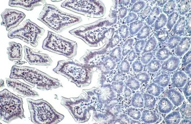
Histone H3K27me3 (trimethyl Lys27) antibody detects Histone H3K27me3 (trimethyl Lys27) protein at nucleus on mouse duodenum by immunohistochemical analysis.
Sample: Paraffin-embedded mouse duodenum.
Histone H3K27me3 (trimethyl Lys27) antibody (GTX121184) diluted at 1:500.
Antigen Retrieval: Trilogy™ (EDTA based, pH 8.0) buffer, 15min

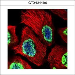
Histone H3K27me3 (Tri-methyl Lys27) antibody detects Histone H3K27me3 (Tri-methyl Lys27) protein at nucleus by immunofluorescent analysis.
Sample: 293T cells were fixed in 4% PFA at RT for 15 min.
Green: Histone H3K27me3 (Tri-methyl Lys27) stained by Histone H3K27me3 (Tri-methyl Lys27) antibody (GTX121184) diluted at 1:500.
Red: alpha Tubulin, a cytoskeleton marker, stained by alpha Tubulin antibody [GT114] (GTX628802) diluted at 1:1000.

Histone H3K27me3 (trimethyl Lys27) antibody detects Histone H3K27me3 (trimethyl Lys27) protein on zebrafish by whole mount immunohistochemical analysis. Sample: PFA-fixed 2 day-post-fertilization zebrafish embryo.Histone H3K27me3 (trimethyl Lys27) antibody (GTX121184) dilution: 1:100.
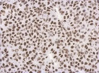
Immunohistochemical analysis of paraffin-embedded Hela xenograft, using Histon H3 (tri-Methyl K27)(GTX121184) antibody at 1:500 dilution.
Antigen Retrieval: Trilogy™ (EDTA based, pH 8.0) buffer, 15min

Histone H3K27me3 (Tri-methyl Lys27) antibody detects Histone H3K27me3 (Tri-methyl Lys27) protein at nucleus by immunofluorescent analysis.Sample: 293T cells were fixed in 4% PFA at RT for 15 min.Green: Histone H3K27me3 (Tri-methyl Lys27) stained by Histone H3K27me3 (Tri-methyl Lys27) antibody (GTX121184) diluted at 1:500.Red: alpha Tubulin, a cytoskeleton marker, stained by alpha Tubulin antibody [GT114] (GTX628802) diluted at 1:1000.
Confocal immunofluorescence analysis (Olympus FV10i) of PFA-fixed A431, using Histone H3 (tri-Methyl K27)(GTX121184) antibody (Green) at 1:500 dilution. Alpha-tubulin filaments were labeled with GTX11304 (Red) at 1:2000.
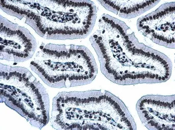
Histone H3K27me3 (Tri-methyl Lys27) antibody detects Histone H3K27me3 (Tri-methyl Lys27) protein at nucleus by immunohistochemical analysis.Sample: Paraffin-embedded mouse intestine.Histone H3K27me3 (Tri-methyl Lys27) stained by Histone H3K27me3 (Tri-methyl Lys27) antibody (GTX121184) diluted at 1:500.Antigen Retrieval: Citrate buffer, pH 6.0, 15 min
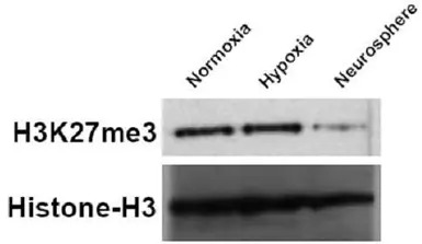
U87 cells were grown under normoxic (DMEM 10% FBS, 16% O2), hypoxic (DMEM 10% FBS, 1% O2), and neurosphere conditions (DMEM/F12, B-27 supplement, growth factor (10ng/ml FGF and 20ng/ml EGF)). Cell lysate were Western blotted for H3K27me3 and Histone-H3 (loading control). The HRP-conjugated anti-rabbit IgG antibody (GTX213110-01) was used to detect the primary antibody.
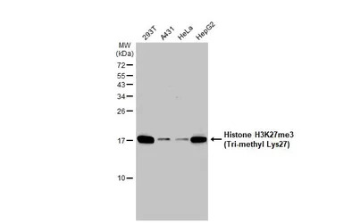
Various whole cell extracts (30 μg) were separated by 15% SDS-PAGE, and the membrane was blotted with Histone H3K27me3 (Tri-methyl Lys27) antibody (GTX121184) diluted at 1:1000. The HRP-conjugated anti-rabbit IgG antibody (GTX213110-01) was used to detect the primary antibody, and the signal was developed with Trident ECL plus-Enhanced.


深圳欣博盛生物科技有限公司
品牌商实名认证
钻石会员
入驻年限:14年
