产品详情
文献和实验
相关推荐
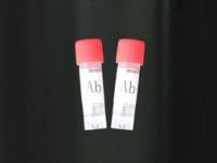
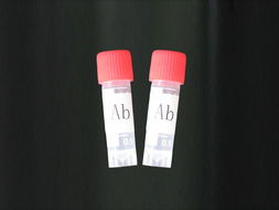
抗体名 :磷酸化细胞信号转导分子SMAD3抗体图片
抗体英文名 :Anti-phospho-Smad3 (Ser204)
靶点 :详见说明书
浓度 :1mg/1ml
应用范围 :产品应用 WB=1:100-500 ELISA=1:500-1000 IP=1:20-100 IHC-P=1:100-500 IHC-F=1:100-500 ICC=1:100-500 IF=1:100-500
宿主 :详见说明书
供应商 :上海一研
库存 :47
级别 :详见说明书
目录编号 :详见说明书
抗原来源 :Rabbit
保质期 :详见说明书
适应物种 :详见说明书
标记物 :详见说明书
克隆性 :多克隆
保存条件 :Store at -20 °C
形态 :详见说明书
亚型 :IgG
免疫原 :KLH conjugated Synthesised phosphopeptide derived from human Smad3 around the phosphorylation site of Ser204 [AG(p-S)PN]
规格 :0.1ml/100μg
公司专业供应的抗体,磷酸化细胞信号转导分子SMAD3抗体图片是用于化学反应、分析化验、研究实验、教学实 验、化学配方使用的纯净化学品,产品品质卓越,价格实惠,多种规格供应,售后完善。英文名称 Anti-phospho-Smad3 (Ser204)
中文名称 磷酸化细胞信号转导分子SMAD3抗体图片
别 名 别 名 SMAD3(phospho S204); SMAD3(phospho Ser204); p-Smad3 (Ser204); hMAD 3; hSMAD3; HSPC193; JV15 2; JV152; MAD (mothers against decapentaplegic Drosophila) homolog 3; MAD3; MADH 3; MADH3; Mothers against decapentaplegic homolog 3; Mothers against DPP homolog 3; SMA and MAD related protein 3; SMAD 3; SMAD; SMAD-3; SMAD3_HUMAN.

浓 度 1mg/1ml
规 格 0.1ml/100μg
抗体来源 Rabbit
克隆类型 polyclonal
交叉反应 Human, Mouse, Rat, Pig, Cow, Horse, Rabbit, Sheep
产品类型 一抗 磷酸化抗体
研究领域 肿瘤 细胞生物 免疫学 信号转导 干细胞 细胞凋亡 转录调节因子
蛋白分子量 predicted molecular weight:
47kDa
性 状 Lyophilized or Liquid
免 疫 原 KLH conjugated Synthesised phosphopeptide derived from human Smad3 around the phosphorylation site of Ser204 [AG(p-S)PN]
亚 型 IgG
纯化方法 affinity purified by Protein A
储 存 液 0.01M PBS, pH 7.4 with 10 mg/ml BSA and 0.1% Sodium azide
产品应用 WB=1:100-500 ELISA=1:500-1000 IP=1:20-100 IHC-P=1:100-500 IHC-F=1:100-500 ICC=1:100-500 IF=1:100-500
(石蜡切片需做抗原修复)
not yet tested in other applications.
optimal dilutions/concentrations should be determined by the end user.
保存条件 Store at -20 °C for one year. Avoid repeated freeze/thaw cycles. The lyophilized antibody is stable at room temperature for at least one month and for greater than a year when kept at -20°C. When reconstituted in sterile pH 7.4 0.01M PBS or diluent of antibody the antibody is stable for at least two weeks at 2-4 °C.
Important Note This product as supplied is intended for research use only, not for use in human, therapeutic or diagnostic applications.
产品介绍 Smad3 is a 50 kDa member of a family of proteins that act as key mediators of TGF beta superfamily signaling in cell proliferation, differentiation and development. The Smad family is divided into three subclasses:
receptor regulated Smads, activin/TGF beta receptor regulated (Smad2 and 3) or BMP receptor regulated (Smad 1, 5, and 8); the common partner, (Smad4) that functions via its interaction to the various Smads; and the inhibitory Smads, (Smad6 and 7). Activated Smad3 oligomerizes with Smad4 upon TGF beta stimulation and translocates as a complex into the nucleus, allowing its binding to DNA and transcription factors. Phosphorylation of the two TGF beta dependent serines 423 and 425 in the C terminus of Smad3 is critical for Smad3 transcriptional activity and TGF beta signaling.
Function :
Receptor-regulated SMAD (R-SMAD) that is an intracellular signal transducer and transcriptional modulator activated by TGF-beta (transforming growth factor) and activin type 1 receptor kinases. Binds the TRE element in the promoter region of many genes that are regulated by TGF-beta and, on formation of the SMAD3/SMAD4 complex, activates transcription. Also can form a SMAD3/SMAD4/JUN/FOS complex at the AP-1/SMAD site to regulate TGF-beta-mediated transcription. Has an inhibitory effect on wound healing probably by modulating both growth and migration of primary keratinocytes and by altering the TGF-mediated chemotaxis of monocytes. This effect on wound healing appears to be hormone-sensitive. Regulator of chondrogenesis and osteogenesis and inhibits early healing of bone fractures (By similarity). Positively regulates PDPK1 kinase activity by stimulating its dissociation from the 14-3-3 protein YWHAQ which acts as a negative regulator.
Subunit :
Monomer; in the absence of TGF-beta. Homooligomer; in the presence of TGF-beta. Heterotrimer; forms a heterotrimer in the presence of TGF-beta consisting of two molecules of C-terminally phosphorylated SMAD2 or SMAD3 and one of SMAD4 to form the transcriptionally active SMAD2/SMAD3-SMAD4 complex. Interacts with TGFBR1. Part of a complex consisting of AIP1, ACVR2A, ACVR1B and SMAD3. Interacts with AIP1, TGFB1I1, TTRAP, FOXL2, PML, PRDM16, HGS and WWP1. Interacts (via MH2 domain) with CITED2 (via C-terminus) (By similarity). Interacts with NEDD4L; the interaction requires TGF-beta stimulation (By similarity). Interacts (via the MH2 domain) with ZFYVE9. Interacts with HDAC1, VDR, TGIF and TGIF2, RUNX3, CREBBP, SKOR1, SKOR2, SNON, ATF2, SMURF2 and TGFB1I1. Interacts with DACH1; the interaction inhibits the TGF-beta signaling. Forms a complex with SMAD2 and TRIM33 upon addition of TGF-beta. Found in a complex with SMAD3, RAN and XPO4. Interacts in the complex directly with XPO4. Interacts (via the MH2 domain) with LEMD3; the interaction represses SMAD3 transcriptional activity through preventing the formation of the heteromeric complex with SMAD4 and translocation to the nucleus. Interacts with RBPMS. Interacts (via MH2 domain) with MECOM. Interacts with WWTR1 (via its coiled-coil domain). Interacts (via the linker region) with EP300 (C-terminal); the interaction promotes SMAD3 acetylation and is enhanced by TGF-beta phosphorylation in the C-terminal of SMAD3. This interaction can be blocked by competitive binding of adenovirus oncoprotein E1A to the same C-terminal site on EP300, which then results in partially inhibited SMAD3/SMAD4 transcriptional activity. Interacts with SKI; the interaction represses SMAD3 transcriptional activity. Component of the multimeric complex SMAD3/SMAD4/JUN/FOS which forms at the AP1 promoter site; required for syngernistic transcriptional activity in response to TGF-beta. Interacts (via an N-terminal domain) with JUN (via its basic DNA binding and leucine zipper domains); this interaction is essential for DNA binding and cooperative transcriptional activity in response to TGF-beta. Interacts with PPM1A; the interaction dephosphorylates SMAD3 in the C-terminal SXS motif leading to disruption of the SMAD2/3-SMAD4 complex, nuclear export and termination of TGF-beta signaling. Interacts (dephosphorylated form via the MH1 and MH2 domains) with RANBP3 (via its C-terminal R domain); the interaction results in the export of dephosphorylated SMAD3 out of the nucleus and termination of the TGF-beta signaling. Interacts with MEN1. Interacts with IL1F7. Interaction with CSNK1G2. Interacts with PDPK1 (via PH domain).

磷酸化细胞信号转导分子SMAD3抗体图片几种抗体概述:
1.抗角蛋白抗体(AKA),又称为抗丝集蛋白抗体或抗角质层抗体。AKA是类风湿关节炎早期诊断和判断预后的指标之一,其对RA患者的诊断的特异性为94% ,而其敏感性为47% ,并是一种鉴别RA和与多发性关节炎相关的丙型肝炎患者的有效检验标记物。
2.抗核周因子( APF), 是定位于口腔黏附膜细胞胞质内的颗粒性蛋白复合物。可在RA患者的血清和关节液中测出,与性别、年龄无关。APF显示了较强的特异性(73%~99%)和敏感性。用间接免疫荧光法检测,方法精密度较差。
3.抗环瓜氨酸肽抗体(CCP),具有相对好的敏感度和特异性。其实,抗核周因子和抗角蛋白抗体,与CCP在化学结构上具有相关性,它们的表位都含有瓜氨酸,称为瓜氨酸相关自身免疫系统。
实验原理 :
(1)特异性结合抗原:抗体本身不能直接溶解或杀伤带有特异抗原的靶细胞,通常需要补体或吞噬细胞等共同发挥效应以清除病原微生物或导致病理损伤。然而,抗体可通过与病毒或毒素的特异性结合,直接发挥中和病毒的作用。
(2)活补体:IgM、IgG1、IgG2和IgG3可通过经典途径激活补体,凝聚的IgA、IgG4和IgE可通过替代途径激活补体。
(3)结合细胞:不同类别的免疫球蛋白,可结合不同种的细胞,参与免疫应答。
(4)可通过胎盘及粘膜:免疫球蛋白G(IgG)能通过胎盘进入胎儿血流中,使胎儿形成自然被动
免疫。免疫球蛋白A(IgA)可通过消化道及呼吸道粘膜,是粘膜局部抗感染免疫的主要因素。
(5)具有抗原性:抗体分子是一种蛋白质,也具有刺机体产生免疫应答的性能。不同的免疫球蛋白分子,各具有不同的抗原性。
(6)抗体对理化因子的抵抗力与一般球蛋白相同:不耐热,60~70℃即被破坏。各种酶及能使蛋白质凝固变性的物质,均能破坏抗体的作用。抗体可被中性盐类沉淀。在上常可用硫酸铵或硫酸钠从免疫血清中沉淀出含有抗体的球蛋白,再经透析法将其纯化。

CL-0338G1(发绿色荧光的小鼠胚胎干细胞)5×10⁶cells/瓶×2
ESAM Others Mouse 小鼠 ESAM / Endothelial Cell Adhesion Molecule 人细胞裂解液 (阳性对照)
小鼠肺大动脉内皮细胞完全培养基 100mL
Eca-109人食管癌细胞 Human esophageal cancer cell line Eca-109. 1640+10% FBS
IL1B Protein Rat 重组大鼠 IL-1 beta / IL1B 蛋白 (mature form)
NCI-H524(人非小细胞肺癌细胞) 5×10⁶cells/瓶×2 HUVEC(人脐静脉血管内皮细胞)
磷酸化细胞信号转导分子SMAD3抗体图片Ana-1(小鼠巨噬细胞) 5×10⁶cells/瓶×2
Anglne(人癌细胞) 5×10⁶cells/瓶×2
AR42J(大鼠胰腺外分泌细胞) 5×10⁶cells/瓶×2
ARPE-19(人视网膜上皮细胞) 5×10⁶cells/瓶×2

上海一研生物科技有限公司
实名认证
钻石会员
入驻年限:8年


