SBI, CYTO120-VA-1, pCT-CD63-GFP (pCMV, Exosome/Secretory, CD63 Tetraspanin Tag), >2 x 10^6 IFUs
价格: 询价
品牌:SBI
 收藏
收藏
产品详情
文献和实验
相关推荐
品牌 :SBI
货号 :CYTO120-VA-1
供应商 :上海觅拓生物
规格 :>2 x 10^6 IFUs
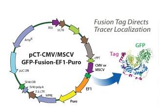
在真核细胞中分子的转运是一个动态过程,Cyto-Tracer提供一种能力,照亮细胞的组成部分并监视其活动,同时定位细胞器,并跟踪细胞内吞作用和细胞外吐作用。SBI创造了一条基于慢病毒载体的Cyto-Tracer应用于长期和深入的实验,其中运用GFP融合蛋白来标记细胞组件,细胞器,小泡和架构。Cyto-Tracer可以用于转染,以及包装进病毒以在原代细胞、肿瘤细胞和干细胞中生产出稳定的GFP跟踪细胞系。亮点:
- 基于慢病毒的细胞跟踪系统
- 对与别的蛋白质进行共同定位的研究很理想
- 实时监控蛋白质的转运
- 有为细胞器、小泡和其它组分构建的质粒
- 明亮的对光稳定的GFP对细胞没有干扰作用。
靶定位:Exosome/Secretory
标记物靶肽:CD63 Tetraspanin


Supporting Data
See some of our exosome Cyto-Tracers in action
The following figure and videos are from:
Garcia NA, et al. Glucose Starvation in Cardiomyocytes Enhances Exosome Secretion and Promotes Angiogenesis in Endothelial Cells. PLoS ONE. 2015. 10(9). PMCID: PMC4578916.
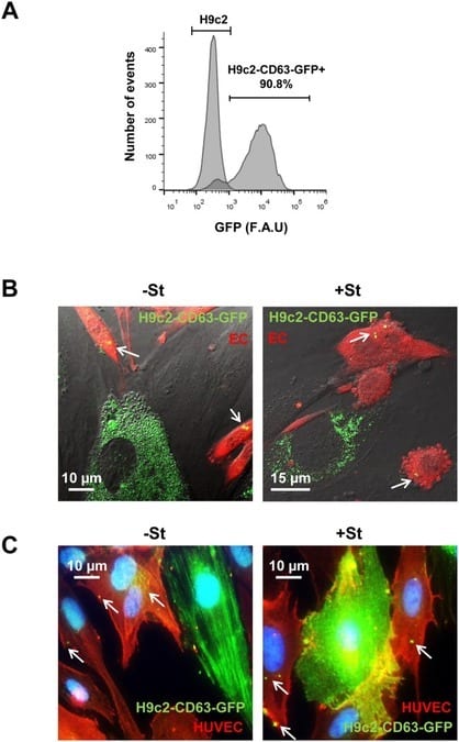
Figure 4 from Garcia, et al. Exosome transfer from CMs to ECs. (A) H9c2 transfected with pCT-CD63-GFP. FACS analysis of 90% GFP positive cells. (B) Representative images from confocal time-lapse microscopy of mouse ECs (ACTB-DsRed) (EC; red) co-cultured with H9C2-CD63-GFP cells (green), previously cultured for 24 h in +/-St medium. Exosome transfer from H9C2 CMs to EC can be observed (S1 and S2 Movies, below). White arrows show CD63-GFP structures inside ECs (C) Representative immunostaining of H9C2-CD63-GFP and HUVEC co-cultures; anti-GFP (green) and anti-CD31 (red). The images illustrate GFP fluorescence from CD63-GFP exosomes in CD31-positive cells (red) after 24 h incubation in +/-St medium. White arrows show CD63-GFP structures inside ECs.
Video S1 from Garcia, et al. Exosome transfer from H9C2-CD63-GFP (green) to endothelial DsRed cells (red) under +St conditions.
Video S2 from Garcia, et al. Exosome transfer from H9C2-CD63-GFP (green) to endothelial DsRed cells (red) under -St conditions.
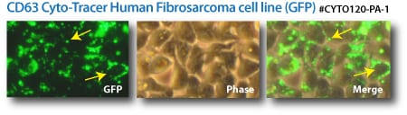
Labeled exosomes. (Top panels) CD63-GFP Cyto-Tracers transfected into a human fibrosarcoma cell line. (Bottom panels) CD9-GFP and CD9-RFP Cyto-Tracers co-transfected into HEK293 cells.


上海觅拓生物科技有限公司
实名认证
金牌会员
入驻年限:9年
