
产品详情
文献和实验
相关推荐
供应商 :江西江蓝纯生物试剂有限公司
库存 :大量
克隆性 :多克隆
保质期 :1年
抗体英文名 :Anti-MAP3K7 antibody
宿主 :Rabbit
浓度 :0.3 mg/ml
保存条件 :-20°C
规格 :25 μl/100 μl/200 μl

Anti-MAP3K7 antibody
Cat. No. JLC223285
Package 25 μl/100 μl/200 μl
Storage -20°C, pH7.4 PBS, 0.05% NaN3, 40% Glycerol
Product overview
Description Anti-MAP3K7 rabbit polyclonal antibody
Applications ELISA, WB, IHC
Immunogen Fusion protein of human MAP3K7
Reactivity Human, Mouse, Rat
Content 0.3 mg/ml
Host species Rabbit
Ig class Immunogen-specific rabbit IgG
Purification Antigen affinity purification
Target information
Symbol MAP3K7
Full name mitogen-activated protein kinase kinase kinase 7
Synonyms TAK1; MEKK7; TGF1a
Swissprot O43318
Target Background
The protein encoded by this gene is a member of the serine/threonine protein kinase family. This kinase mediates the signaling transduction induced by TGF beta and morphogenetic protein (BMP), and controls a variety of cell functions including transcription regulation and apoptosis. In response to IL-1, this protein forms a kinase complex including TRAF6, MAP3K7P1/TAB1 and MAP3K7P2/TAB2; this complex is required for the activation of nuclear factor kappa B. This kinase can also activate MAPK8/JNK, MAP2K4/MKK4, and thus plays a role in the cell response to environmental stresses. Four alternatively spliced transcript variants encoding distinct isoforms have been reported.
Applications
Immunohistochemistry
Predicted cell location: Cytoplasm and Cell membrane
Positive control: Human esophagus cancer
Recommended dilution: 10-50 Predicted cell location: Cytoplasm and Cell membrane
Positive control: Human lung cancer
Recommended dilution: 10-50
The image on the left is immunohistochemistry of paraffin-embedded Human esophagus cancer tissue using ml223285(MAP3K7 Antibody) at dilution 1/20, on the right is treated with fusion protein. (Original magnification: ×200) The image on the left is immunohistochemistry of paraffin-embedded Human lung cancer tissue using ml223285(MAP3K7 Antibody) at dilution 1/20, on the right is treated with fusion protein. (Original magnification: ×200)
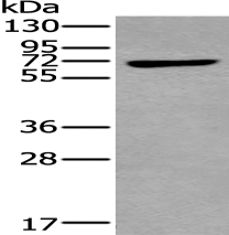
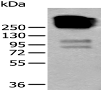
Western blotting
Predicted band size:67 kDa
Positive control:HEPG2 cell lysate
Recommended dilution: 500-2000
Gel: 8%SDS-PAGE
Lysate: 40 μg
Lane: HEPG2 cell lysate
Primary antibody: ml223285(MAP3K7 Antibody) at dilution 1/250
Secondary antibody: Goat anti rabbit IgG at 1/8000 dilution
Exposure time: 1 minute
ELISA
Recommended dilution: 2000-5000


江西江蓝纯生物试剂有限公司
实名认证
钻石会员
入驻年限:8年
