
产品详情
文献和实验
相关推荐


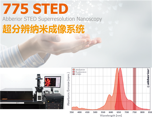
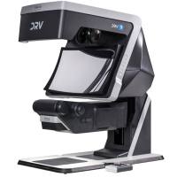
受激辐射光淬灭超分辨率共聚焦显微影像系统 (pulsed-STED), 源自诺贝尔奖团队的设计, 随时拥有先进的技术以因应未来各种应用. 也是唯一具备可以客制化的灵活系统, 应用空间宽广无限, 同时, 拥有最高解析的超分辨率.
Abberior 的使命即是开发更实用的超分辨技术, 发挥超分辨的效能, 从光学解析的持续突破, 落实到单一纳米级的成像解析, 与, 活细胞的动态超分辨成像, 扩大样本厚度的深层取像, 引领科研的深度成就, 落实用户的仪器使用价值 !

Abberior 775 STED 具有下列优势 :


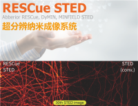
2-color STED image with negligible crosstalk taken with our 2-color package Abberior STAR 580/STAR 600 and Abberior STAR 635P.. Comparison of a 2-color confocal (left) and the corresponding STED (right) microscopy image; overview (top panels), close-up (bottom panels). Labels are Abberior STAR 580 (Nup, green) and STAR 635P (lamin, purple).
Abberior 775 STED 基于共聚焦扫描成像的脉冲式超分辨, 可以高速一次性成像, 获得纯光学解析的高质量影像, 无须耗时耗力的虚拟图像函数运算 ( 其他技术, 随机光学重建技术, 如, STORM, PALM, dSTORM/GSD, 都需要仰赖长时间的虚拟图像运算 ), 所以, 在 2D/3D/4D 的多色荧光成像应用, 可以快速轻松取得. 无论在解析与速度, 都是其他随机光学重建技术所无法达到的 !
主动式自动对焦系统 (Autofocus-STED)
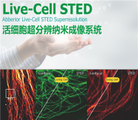
特别推荐, 主动式自动对焦系统 ( Autofocus-STED ), 独家的前瞻性设计, 也是全球唯一. 在超分辨成像的过程中, 随时保持聚焦, 适合活动中的活细胞或其他动态样本成像应用. 此对焦设计, 也落实了超分辨在活细胞成像应用的关键因素之一.


Live-cell Confocal and Live-cell STED images of SiR tubulin labelled mammalian cells; In total ~1.5h (100 frames)
Abberior 775 STED 可以依照用户需求, 配合实验规划, 染剂应用, 提供最佳的超分辨组配


| 超分辨成像扫描 |
|
|
| Pulsed STED - 脉冲式超分辨技术 |
|
|
| Easy3D STED 专利技术 |
|
|
| 脉冲式STED 压制激光 (标配) |
|
|
| 脉冲式STED 压制激光 (选配) |
|
|
| 共聚焦激光 |
|
|
| 侦测器 |
|
|
| 2D (XY) 光学解析 |
|
|
| 3D (XYZ) 光学解析 |
|
|
| Z-轴 (Z) 光学解析 |
|
|
| 平台设计 |
|
|
| 智能光照调控 |
|
|
| 荧光标定染剂, 荧光蛋白 |
|
|


Adaptive optics easy3D STED enables deep imaging of cleared adult kidney samples
Comparison of a XZ-slice deep into a renal corpuscle using conventional 3D STED imaging and an easy3D STED imaging. easy3D allowed imaging up to 80 µm into cleared rat kidney tissue using an oil objective. Without adaptive optics, the mismatch between immersion oil and sample leads to complete signal loss.
Labels: Nephrin (red, Abberior STAR635P) and Podocin (green, AlexaFluor594). Sample was prepared by D. Unnersjö Jess and H.G. Blom @ KTH Stockholm, Sweden.

| STED光路设计 |
Abberior 专利技术 |
已淘汰的传统技术 |
| STED 技術 |
脉冲式 Pulsed STED |
CW STED, g-STED |
| 光路设计 |
单一 STED 光路 (2D — 3D) 产生最佳同心圆光斑, 提供最高超分辨光学解析SLM 技术, 光斑可调, 像差修正 |
|
| 光路稳定性 |
极为稳定 因为单一光路, 稳定性极高. 调整也容易. 不受温湿度影响 |
|
| 使用方法 |
简易, 快速 |
|
| 2D (XY) 解析 |
< 20 nm 已发展达到 XY 单一纳米级的分辨力 |
|
| 3D (XYZ) 解析 |
已发展达到 XYZ < 70 nm 分辨力 |
|
| 3D STED 成像深度(厚度) |
> 80 μm, 成像深度(厚度) 已达 120 微米 |
|
| 是否可使用更种物镜 ? |
Oil, Water, Glycerin, Silicon oil, Dipping len (镜头的像差修正) |
|

Abberior 775 STED 在 3D STED 成像, 使用水镜或甘油镜, 皆可做像差的修正, 得到更高解析更清新的厚度成像.
J. Heine, C. A. Wurm, J. Keller-Findeisen, A. Schönle, B. Harke, M. Reuss, F. R. Winter, G. Donnert, "Three dimensional live-cell STED microscopy at increased depth using a water immersion objective", Review of Scientific Instruments (2018)

Shown is an image of a renal corpuscle showing Nephrin (red, Abberior STAR635P) structures inbetween the Podocin slits (green, AlexaFluor594). Sample was prepared by D. Unnersjö Jess and H.G. Blom @ KTH Stockholm, Sweden.

德国Abberior来自诺贝尔奖得主 Stefan Hell 的科研团队, 是全球唯一同时拥有最尖端的超分辨技术, 超分辨仪器, 超分辨染剂, 超分辨研发团队, 超分辨科研团队., 也是引领全球超分辨技术与科研应用的领航者.
Abberior 希望用户的每一平台, 都可以在拥有的第一刻起, 永远可以导入最新开发的技术与设备, 可以无限制的因应未来各种应用需求. 所以, 每一套 Abberior STED 系统, 都具有无限的扩充功能, 可以导入所有 Abberior 新开发的各种技术与设备.
Abberior 775 STED 是最尖端与标准的超分辨纳米成像系统. 从超高的光学分辨力, 2D 解析达 20 nm, 甚至直逼到单一纳米的解析, 从 3D 解析达 70x70x70 nm 的分辨力, 直逼更深层的超分辨纳米成像. 所以, 基本应用可依导入的光学组件, 落实下列的基础应用 :
- 超高解析的 2D, 3D, 4D 超分辨纳米荧光成像 (Pulsed STED Superresolution)
- 多色超分辨纳米荧光成像 (Multi-color STED)
- 高速活细胞影像 (Live-Cell STED)
- 深层的超分辨纳米荧光成像 (2-Photon STED)
- 超分辨分子动态成像分析 (STED-FLIM, FCS, FRET)


- Abberior ImSpector 导航式图像控制分析软件, 简易使用, 人人都可轻易上手.
- 可以控制用户的外部组件 (独家唯一的开放式设计, 可以提升仪器使用效益, 可以提升仪器的扩充功能)
- 内建超分辨与共聚焦专用 deconvolution 分析软件
- 可以兼容第三方分析软件, 如 Matlab, Python 数据控制, Amira, Imaris, Image Surfer2, ParaVeiw 3D-分析软件, Fiji, Cell Profiler, Icy 数据分析


Abberior 公司是唯一具有先进的超分辨技术, 仪器, 染剂, 研发团队, 科研团队.
- 两大系列荧光染剂 LIVE (别针对活细胞染色应用) 及 STAR 系列染剂. 特别针对 775 nm 及 595 nm STED 所适用. (也可针对 592/660 激光).
- 新型荧光分子团, 及耐受高强度激光, 可长时间使用, 不易荧光漂白, 具有高亮度(高QE)特性, 可产生亮丽的高解析荧光成像.
- 可避免样本光毒害, 保护实验样本.
- 良好的水溶性, 具有优越的膜透性, 常规染色应用于 肌动蛋白, 维管蛋白, 核酸, 细胞核, 溶酶体, 细胞骨架. 包括多色荧光, 具有超分辨专属的有机荧光染剂, 应用于活细胞成像
- 多功能染剂衍生物,可选择自行接任何抗体. 可替换Alexa, ATTO或者Cy染剂在Confocal或STED上的应用

范例 : 双色 STED 荧光应用 (775 nm STED), 可选用 STAR 520XP, STAR 635P, 可再配合 SiR 染剂.

2C-Pack STED 775 IR -LS-
- Abberior STAR 雙色染劑包 (適用 775 STED : 2-color STED images with the Abberior STAR 520SXP and Abberior STAR635P).
- 2C packs using a regular dye in the red spectra region together with a long stokes shift dye.
- The packages contain two phosphorylated dyes - Abberior STAR 470SXP and Abberior STAR 635P or Abberior STAR 520SXP and Abberior STAR 635P,. Abberior STAR 635P is our flagship dye for STED microscopy in the infrared spectral region (Exc. 620 - 650 nm/ STED 750 - 780 nm). Abberior STAR 470SXP (Exc. 450 - 490 nm) and Abberior STAR 520SXP (Exc. 500 - 540 nm) are long-stokes shift partners for 2-color STED with one STED laser at ~750nm - 775nm. The dye pair is tested and verified for the Abberior 2C STED 775 microscope ..
Abberior LIVE 580 活细胞超分辨成像应用的染剂

live-cell dyes. The striking feature of this dye series is their cell permeability, enabling live-cell experiments. The LIVE dye series offers unique labeling options - live-cell imaging with organic dyes now becomes possible! The LIVE dyes combine the best of two worlds:
- organic dyes with the highest brightness & photostability among all fluorescent markers, and
- live-cell labeling out of the box.
Confocal (left) and super resolution (right) microscopy image of tubulin stained with LIVE 580 tubulin (cabazitaxel) in living human fibroblasts. The image was taken with an Abberior Instruments STED microscope.


Source: Göttfert, F., C. A. Wurm, V. Mueller, S. Berning, V. C. Cordes, A. Honigmann, S. W. Hell:
Coaligned Dual-Channel STED Nanoscopy and Molecular Diffusion Analysis at 20 nm Resolution" Biophys. J. 105, L01 - L03
Red: gp210 / Green: Several proteins in the central channel
RAW : 原始图文件, 未经任何图像修饰. ( 唯有 Abberior STED 具有一次性共聚焦超分辨扫描, 直接成像取得纯光学解析的高解析影像, 无须耗时耗力的图像处理)

Confocal and 2D Pulsed STED ( Abberior 775 STED ) imaging of dendrites in brain slices. GFP-tagged proteins were expressed and immunolabelled using primary antibodies against GFP and secondary antibodies coupled to Abberior STAR 635P. Sample was prepared by O. Kaplan and H. Kawabe @ MPI of Experimental Medicine, Göttingen, Germany.

Growth cone at the tip of the axon of a primary hippocampal neuron at 1 day in vitro. Microtubules (Tuj1 labelling) are bundled in the central-domain suggesting a pausing state. The molecular motor myosin IIB (confocal) is enriched at the transition-zone, along the F-actin arcs. In the peripheral domain actin forms bundles in the filopodia. Imaged with Abberior Expert Line - 775 STED @ MPIbpc by Elisa D’Este.

-
-
J. Hanne, F. Goettfert, J. Schimer, M. Anders-Oesswein, J. Konvalinka, J. Engelhardt, B. Mueller, S. W. Hell, H.-G. Kraeusslich "Stimulated Emission Depletion Nanoscopy Reveals Time-Course of Human Immunodeficiency Virus Proteolytic Maturation", ACS Nano (2016)
-
O. Steshenko, D. M. Andrade, A. Honigmann, V. Mueller, F. Schneider, E. Sezgin, S. W. Hell, M. Simons, C. Eggeling, "Reorganization of Lipid Diffusion by Myelin Basic Protein as Revealed by STED Nanoscopy", Biophysical Journal 110, 2441–2450, Jun. (2016)
-
H. Batoulis, T. H. Schmidt, P. Weber, J.-G. Schloetel, C. Kandt, T. Lang, "Concentration Dependent Ion-Protein Interaction Patterns Underlying Oligomerization Behaviours", Scientific Reports, Apr. (2016)
-
K. Kehrein, R. Schilling, B. V. Möller-Hergt, C. A. Wurm, S. Jakobs, T. Lamkemeyer, T. Langer, M. Ott, "Organization of Mitochondrial Gene Expression in Two Distinct Ribosome-Containing Assemblies", Cell Rep., Feb. 12, pii: S2211-1247(15)00025-X (2015)
-
M. P. Clausen, S. Galiani, J. Bernardino de la Serna, M. Fritsche, J. Chojnacki, K. Gehmlich, B. C. Lagerholm, C. Eggeling,"Pathways to optical STED microscopy", Nanobioimaging 1-12, (2013)
-
G. Vicidomini, G. Moneron, K. Y. Han, V. Westphal, H. Ta, M. Reuss, J. Engelhardt, C. Eggeling, S. W. Hell, "Sharper low-power STED nanoscopy by time gating", Nature Meth. 8, 571 - 573. (2011)
-
B. Harke, C. K. Ullal, J. Keller, S. W. Hell, "Three-Dimensional Nanoscopy of Colloidal Crystals", Nano Lett. 8, 1309 (2008)
-
L. Große, C. A. Wurm, C. Brüser, D. Neumann, D. C. Jans, S. Jakobs, "Bax assembles into large ring-like structures remodeling the mitochondrial outer membrane in apoptosis", EMBO J., (2016)
-
K. Kolmakov, C. A. Wurm, D. N. H. Meineke, F. Göttfert, V. P. Boyarskiy, V. N. Belov, S. W. Hell, "Polar Red-Emitting Rhodamine Dyes with Reactive Groups: Synthesis, Photophysical Properties, and Two-Color STED Nanoscopy Applications", Chem. Eur. J. 20, 146 - 157 (2014)
-
F. Göttfert, C. A. Wurm, V. Mueller, S. Berning, V. C. Cordes, A. Honigmann, S. W. Hell, "Coaligned Dual-Channel STED Nanoscopy and Molecular Diffusion Analysis at 20 nm Resolution", Biophysical Journal Volume 105, L01–L03 (2013)
-
C. A. Wurm, K. Kolmakov, F. Göttfert, H. Ta, M. Bossi, H. Schill, S. Berning, S. Jakobs, G. Donnert, V. N. Belov, S. W. Hell, "Novel red fluorophores with superior performance in STED microscopy", Optical Nanoscopy 1, 1 – 7 (2012)
-
K. Kolmakov, C. A. Wurm, R. Hennig, E. Rapp, S. Jakobs, V. N. Belov, S. W. Hell, "Red-Emitting Rhodamines with Hydroxylated, Sulfonated, and Phosphorylated Dye Residues and Their Use in Fluorescence Nanoscopy" Chemistry", A European Journal 18, 12986 - 12998 (2012)
-
B. Harke, J. Keller, C. K. Ullal, V. Westphal, A. Schönle, S. W. Hell, "Resolution scaling in STED microscopy", Opt. Express 16, 4154-4162 (2008)
-
B. Harke, J. V. Chacko, H. Haschke, C. Canale, A. Diaspro, " A novel nanoscopic tool by combining AFM with STED microscopy", Optical Nanoscopy 2012, 1:3
-
J. V. Chacko, B. Harke, C. Canale, A. Diaspro, "Cellular level nanomanipulation using atomic force microscope aided with superresolution imaging", J Biomed Opt. 2014; 19(10):105003.
-
J. V. Chacko, C. Canale, B. Harke, A. Diaspro, "Sub-diffraction nano manipulation using STED AFM", PLoS One. 2013 Jun 17;8(6):e66608
-
F. Göttfert, T. Pleiner, J. Heine, V. Westphal, D. Görlich, S. J. Sahl, S. W. Hell, "Strong signal increase in STED fluorescence microscopy by imaging regions of subdiffraction extent", PNAS 1621495114 (2017)
-


上海宇北医疗器械有限公司
实名认证
钻石会员
入驻年限:7年
