
产品详情
文献和实验
相关推荐
供应商 :上海联迈生物工程有限公司
库存 :大量
目录编号 :LM-7529R
克隆性 :多克隆
抗原来源 :Rabbit
保质期 :1年
抗体英文名 :Collagen VIII alpha 1
抗体名 :8型胶原/内皮胶原蛋白抗体
宿主 :Rabbit
适应物种 :Human, Mouse, Rat, Chicken, Dog, Pig, Cow,
免疫原 :KLH conjugated synthetic peptide derived from human Collagen VIII alpha 1 (672-718aa):641-744/744
亚型 :IgG
形态 :Lyophilized or Liquid
应用范围 :WB=1:500-2000 ELISA=1:500-1000 IHC-P=1:400-800 IHC-F=1:400-800 IF=1:100-500 (石蜡切片需做抗原修复)
浓度 :1mg/ml
保存条件 :Store at -20 °C
规格 :100ul 200ul
Collagen VIII alpha 1 8型胶原/内皮胶原蛋白抗体| 英文名称 | Collagen VIII alpha 1 |
| 中文名称 | 8型胶原/内皮胶原蛋白抗体 |
| 别 名 | C3orf7; CO8A1_HUMAN; COL8A1; Collagen alpha 1(VIII) chain [Precursor]; Endothelial collagen; MGC9568; Vastatin. |
| 规格价格 | 100ul/1380元 购买 200ul/2200元 购买 大包装/询价 |
| 说 明 书 | 100ul 200ul |
| 研究领域 | 心血管 信号转导 糖尿病 细胞骨架 细胞外基质 |
| 抗体来源 | Rabbit |
| 克隆类型 | Polyclonal |
| 交叉反应 | Human, Mouse, Rat, Chicken, Dog, Pig, Cow, |
| 产品应用 | WB=1:500-2000 ELISA=1:500-1000 IHC-P=1:400-800 IHC-F=1:400-800 IF=1:100-500 (石蜡切片需做抗原修复) not yet tested in other applications. optimal dilutions/concentrations should be determined by the end user. |
| 分 子 量 | 19/79kDa |
| 细胞定位 | 分泌型蛋白 |
| 性 状 | Lyophilized or Liquid |
| 浓 度 | 1mg/ml |
| 免 疫 原 | KLH conjugated synthetic peptide derived from human Collagen VIII alpha 1 (672-718aa):641-744/744 |
| 亚 型 | IgG |
| 纯化方法 | affinity purified by Protein A |
| 储 存 液 | 0.01M TBS(pH7.4) with 1% BSA, 0.03% Proclin300 and 50% Glycerol. |
| 保存条件 | Store at -20 °C for one year. Avoid repeated freeze/thaw cycles. The lyophilized antibody is stable at room temperature for at least one month and for greater than a year when kept at -20°C. When reconstituted in sterile pH 7.4 0.01M PBS or diluent of antibody the antibody is stable for at least two weeks at 2-4 °C. |
| PubMed | PubMed |
| 产品介绍 | background: Macromolecular component of the subendothelium. Major component of the Descemet's membrane (basement membrane) of corneal endothelial cells. Also component of the endothelia of blood vessels. Necessary for migration and proliferation of vascular smooth muscle cells and thus, has a potential role in the maintenance of vessel wall integrity and structure, in particular in artherogenesis. Vastatin, the C-terminal fragment comprising the NC1 domain, inhibits aortic endothelial cell proliferation and causes cell apoptosis. Function: Macromolecular component of the subendothelium. Major component of the Descemet's membrane (basement membrane) of corneal endothelial cells. Also component of the endothelia of blood vessels. Necessary for migration and proliferation of vascular smooth muscle cells and thus, has a potential role in the maintenance of vessel wall integrity and structure, in particular in atherogenesis. Vastatin, the C-terminal fragment comprising the NC1 domain, inhibits aortic endothelial cell proliferation and causes cell apoptosis. Subunit: Homodimer; antiparallel disulfide-linked dimer. Heterodimer with PDGFB; antiparallel disulfide-linked dimer. The PDGFA homodimer interacts with PDGFRA homodimers, and with heterodimers formed by PDGFRA and PDGFRB. The heterodimer composed of PDGFA and PDGFB interacts with PDGFRA homodimers, and with heterodimers formed by PDGFRA and PDGFRB. Interacts with CSPG4. Subcellular Location: Secreted, extracellular space, extracellular matrix, basement membrane. Tissue Specificity: Expressed primarily in the subendothelium of large blood vessels. Also expressed in arterioles and venules in muscle, heart, kidney, spleen, umbilical cord, liver and lung and is also found in connective tissue layers around hair follicles, around nerve bundles in muscle, in the dura of the optic nerve, in cornea and sclera, and in the perichondrium of cartilaginous tissues. In the kidney, expressed in mesangial cells, glomerular endothelial cells, and tubular epithelial cells. Also expressed in mast cells, and in astrocytes during the repair process. Expressed in Descemet's membrane. Specifically expressed in peritoneal fibroblasts and mesothelial cells. Post-translational modifications: Prolines at the third position of the tripeptide repeating unit (G-X-Y) are hydroxylated in some or all of the chains. Proteolytically cleaved by neutrophil elastase, in vitro. Proteolytic processing produces the C-terminal NC1 domain fragment, vastatin. Similarity: Belongs to the PDGF/VEGF growth factor family. SWISS: P27658 Gene ID: 1295 Database links: UniProtKB/Swiss-Prot: P27658.2 Important Note: This product as supplied is intended for research use only, not for use in human, therapeutic or diagnostic applications. |
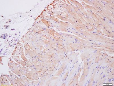
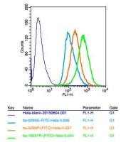
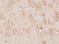
| 产品图片 | 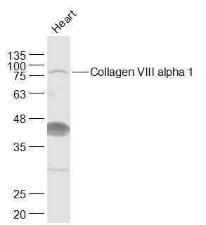 Sample: Heart (Rat) Lysate at 40 ug Primary: Anti-Collagen VIII alpha 1 (bs-7529R) at 1/1000 dilution Secondary: IRDye800CW Goat Anti-Rabbit IgG at 1/20000 dilution Predicted band size: 19/79 kD Observed band size: 79 kD 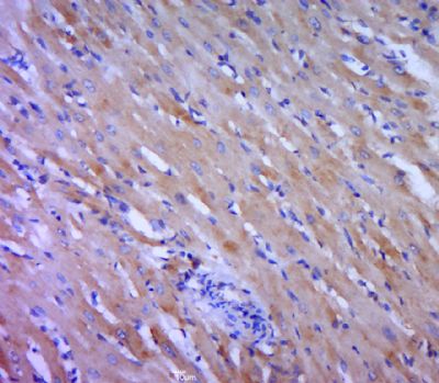 Paraformaldehyde-fixed, paraffin embedded (rat heart tissue); Antigen retrieval by boiling in sodium citrate buffer (pH6.0) for 15min; Block endogenous peroxidase by 3% hydrogen peroxide for 20 minutes; Blocking buffer (normal goat serum) at 37°C for 30min; Antibody incubation with (Collagen VIII alpha 1) Polyclonal Antibody, Unconjugated (bs-7529R) at 1:400 overnight at 4°C, followed by a conjugated secondary (sp-0023) for 20 minutes and DAB staining.  Tissue/cell: smooth muscle of mouse stomach; 4% Paraformaldehyde-fixed and paraffin-embedded; Antigen retrieval: citrate buffer ( 0.01M, pH 6.0 ), Boiling bathing for 15min; Block endogenous peroxidase by 3% Hydrogen peroxide for 30min; Blocking buffer (normal goat serum,C-0005) at 37℃ for 20 min; Incubation: Anti-Collagen VIII alpha 1 Polyclonal Antibody, Unconjugated(bs-7529R) 1:200, overnight at 4°C, followed by conjugation to the secondary antibody(SP-0023) and DAB(C-0010) staining |


上海联迈生物工程有限公司
品牌商实名认证
钻石会员
入驻年限:8年
