
产品详情
文献和实验
相关推荐
供应商 :上海联迈生物工程有限公司
库存 :大量
目录编号 :LM-1447R
克隆性 :多克隆
抗原来源 :Rabbit
保质期 :1年
抗体英文名 :HIF 2 alpha
抗体名 :缺氧诱导因子2α /HIF-2α/HIF-2-alpha抗体
宿主 :Rabbit
适应物种 :Human, Mouse, Rat, Pig, Cow, Horse,
免疫原 :KLH conjugated synthetic peptide derived from mouse HIF 2 Alpha:1-130/874
亚型 :IgG
形态 :Lyophilized or Liquid
应用范围 :WB=1:500-2000 ELISA=1:500-1000 IHC-P=1:400-800 IHC-F=1:400-800 Flow-Cyt=1µg/Test IF=1:100-500 (石蜡切片需做抗原修复)
浓度 :1mg/ml
保存条件 :Store at -20 °C
规格 :50ul 100ul 200ul
HIF 2 alpha缺氧诱导因子2α /HIF-2α/HIF-2-alpha抗体| 英文名称 | HIF 2 alpha |
| 中文名称 | 缺氧诱导因子2α /HIF-2α/HIF-2-alpha抗体 |
| 别 名 | Basic helix loop helix PAS protein MOP2; Basic-helix-loop-helix-PAS protein MOP2; bHLHe73; Class E basic helix-loop-helix protein 73; ECYT4; Endothelial PAS domain containing protein 1; Endothelial pas domain protein 1; Endothelial PAS domain-containing protein 1; EPAS 1; EPAS-1; EPAS1; EPAS1_HUMAN; HIF 1 alpha like factor; HIF 2 alpha; HIF-1-alpha-like factor; HIF-2-alpha; HIF2-alpha; HIF2A; HLF; Hypoxia inducible factor 2 alpha; Hypoxia inducible factor 2 alpha subunit; Hypoxia-inducible factor 2-alpha; Member of PAS protein 2; Member of pas superfamily 2; MOP 2; MOP2; PAS domain-containing protein 2; PASD2. |
| 规格价格 | 50ul/780元 购买 100ul/1380元 购买 200ul/2200元 购买 大包装/询价 |
| 说 明 书 | 50ul 100ul 200ul |
| 研究领域 | 肿瘤 免疫学 信号转导 细胞凋亡 |
| 抗体来源 | Rabbit |
| 克隆类型 | Polyclonal |
| 交叉反应 | Human, Mouse, Rat, Pig, Cow, Horse, |
| 产品应用 | WB=1:500-2000 ELISA=1:500-1000 IHC-P=1:400-800 IHC-F=1:400-800 Flow-Cyt=1µg/Test IF=1:100-500 (石蜡切片需做抗原修复) not yet tested in other applications. optimal dilutions/concentrations should be determined by the end user. |
| 分 子 量 | 96kDa |
| 细胞定位 | 细胞核 细胞浆 |
| 性 状 | Lyophilized or Liquid |
| 浓 度 | 1mg/ml |
| 免 疫 原 | KLH conjugated synthetic peptide derived from mouse HIF 2 Alpha:1-130/874 |
| 亚 型 | IgG |
| 纯化方法 | affinity purified by Protein A |
| 储 存 液 | 0.01M TBS(pH7.4) with 1% BSA, 0.03% Proclin300 and 50% Glycerol. |
| 保存条件 | Store at -20 °C for one year. Avoid repeated freeze/thaw cycles. The lyophilized antibody is stable at room temperature for at least one month and for greater than a year when kept at -20°C. When reconstituted in sterile pH 7.4 0.01M PBS or diluent of antibody the antibody is stable for at least two weeks at 2-4 °C. |
| PubMed | PubMed |
| 产品介绍 | background: Transcription factor involved in the induction of oxygen regulated genes. Binds to core DNA sequence 5'-[AG]CGTG-3' within the hypoxia response element (HRE) of target gene promoters. Regulates the vascular endothelial growth factor (VEGF) expression and seems to be implicated in the development of blood vessels and the tubular system of lung. May also play a role in the formation of the endothelium that gives rise to the blood brain barrier. Potent activator of the Tie-2 tyrosine kinase expression. Activation requires recruitment of transcriptional coactivators such as CREBPB and probably EP300. Interaction with redox regulatory protein APEX seems to activate CTAD. Function: Transcription factor involved in the induction of oxygen regulated genes. Binds to core DNA sequence 5'-[AG]CGTG-3' within the hypoxia response element (HRE) of target gene promoters. Regulates the vascular endothelial growth factor (VEGF) expression and seems to be implicated in the development of blood vessels and the tubular system of lung. May also play a role in the formation of the endothelium that gives rise to the blood brain barrier. Potent activator of the Tie-2 tyrosine kinase expression. Activation requires recruitment of transcriptional coactivators such as CREBPB and probably EP300. Interaction with redox regulatory protein APEX seems to activate CTAD. Subunit: Efficient DNA binding requires dimerization with another bHLH protein. Heterodimerizes with ARNT. Interacts with CREBPB. Subcellular Location: Nucleus (Potential). Tissue Specificity: Expressed in most tissues, with highest levels in lung, followed by heart, kidney, brain and liver. Predominantly expressed in endothelial cells. Also found in smooth muscle cells of the uterus, neurons, and brown adipose tissue. High expression in embryonic choroid plexus and kidney glomeruli. Post-translational modifications: In normoxia, is probably hydroxylated on Pro-405 and Pro-530 by EGLN1/PHD1, EGLN2/PHD2 and/or EGLN3/PHD3. The hydroxylated prolines promote interaction with VHL, initiating rapid ubiquitination and subsequent proteasomal degradation. Under hypoxia, proline hydroxylation is impaired and ubiquitination is attenuated, resulting in stabilization. In normoxia, is hydroxylated on Asn-851 by HIF1AN thus probably abrogating interaction with CREBBP and EP300 and preventing transcriptional activation. Phosphorylated on multiple sites in the CTAD. The iron and 2-oxoglutarate dependent 3-hydroxylation of asparagine is (S) stereospecific within HIF CTAD domains. Similarity: Contains 1 basic helix-loop-helix (bHLH) domain. Contains 1 PAC (PAS-associated C-terminal) domain. Contains 2 PAS (PER-ARNT-SIM) domains. SWISS: P97481 Gene ID: 13819 Database links: Entrez Gene: 282711 Cow Entrez Gene: 2034 Human Entrez Gene: 13819 Mouse Entrez Gene: 100037272 Pig Entrez Gene: 29452 Rat Omim: 603349 Human SwissProt: Q99814 Human SwissProt: P97481 Mouse SwissProt: Q9JHS1 Rat Unigene: 468410 Human Unigene: 1415 Mouse Unigene: 55138 Rat Important Note: This product as supplied is intended for research use only, not for use in human, therapeutic or diagnostic applications. HIF-2α也是机体对缺氧适应性调节中的重要调控蛋白.有学者认为:HIF-2α对肿瘤的能量代谢和新血管生成有促进作用,微血管的形成可抑制肿瘤细胞的凋亡,从而促进肿瘤的恶性发展。 |
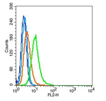
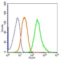
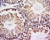
| 产品图片 |  Sample: Lung(Rat) lysate at 30ug; Primary: Rabbit Anti-HIF 2α (bs-1447R) at 1:200 dilution; Secondary: HRP conjugated Goat Anti-Rabbit IgG(bs-0295G-HRP) at 1: 3000 dilution; Predicted band size : 96kD Observed band size : 96kD  Paraformaldehyde-fixed, paraffin embedded (Mouse brain); Antigen retrieval by boiling in sodium citrate buffer (pH6.0) for 15min; Block endogenous peroxidase by 3% hydrogen peroxide for 20 minutes; Blocking buffer (normal goat serum) at 37°C for 30min; Antibody incubation with (HIF 2 alpha) Polyclonal Antibody, Unconjugated (bs-1447R) at 1:500 overnight at 4°C, followed by a conjugated secondary (sp-0023) for 20 minutes and DAB staining.  Tissue/cell: human glioma tissue; 4% Paraformaldehyde-fixed and paraffin-embedded; Antigen retrieval: citrate buffer ( 0.01M, pH 6.0 ), Boiling bathing for 15min; Block endogenous peroxidase by 3% Hydrogen peroxide for 30min; Blocking buffer (normal goat serum,C-0005) at 37℃ for 20 min; Incubation: Anti-HIF 2α Polyclonal Antibody, Unconjugated(bs-1447R) 1:200, overnight at 4°C, followed by conjugation to the secondary antibody(SP-0023) and DAB(C-0010) staining  Tissue/cell: mouse lymph node;4% Paraformaldehyde-fixed and paraffin-embedded; Antigen retrieval: citrate buffer ( 0.01M, pH 6.0 ), Boiling bathing for 15min; Blocking buffer (normal goat serum,C-0005) at 37℃ for 20 min; Incubation: Anti-HIF 2αPolyclonal Antibody, Unconjugated(bs-1447R) 1:200, overnight at 4°C; The secondary antibody was Goat Anti-Rabbit IgG, Cy3 conjugated(bs-0295G-Cy3)used at 1:200 dilution for 40 minutes at 37°C. DAPI(5ug/ml,blue,C-0033) was used to stain the cell nuclei 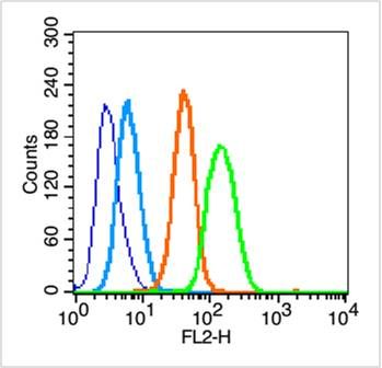 Blank control (blue line): Hep G2 (blue). Primary Antibody (green line): Rabbit Anti-HIF 2 alpha antibody (bs-1447R) Dilution: 1μg /10^6 cells; Isotype Control Antibody (orange line): Rabbit IgG . Secondary Antibody (white blue line): Goat anti-rabbit IgG-PE Dilution: 1μg /test. Protocol The cells were fixed with 70% ethanol (Overnight at 4℃) and then permeabilized with 90% methanol for 20 min at -20℃. Cells stained with Primary Antibody for 30 min at room temperature. The cells were then incubated in 1 X PBS/2%BSA/10% goat serum to block non-specific protein-protein interactions followed by the antibody for 15 min at room temperature. The secondary antibody used for 40 min at room temperature. Acquisition of 20,000 events was performed.  Blank control: RSC96(blue). Primary Antibody:Rabbit Anti-HIF 2α antibody(bs-1447R), Dilution: 1μg in 100 μL 1X PBS containing 0.5% BSA; Isotype Control Antibody: Rabbit IgG(orange) ,used under the same conditions ); Secondary Antibody: Goat anti-rabbit IgG-PE(white blue), Dilution: 1:200 in 1 X PBS containing 0.5% BSA. Protocol The cells were fixed with 2% paraformaldehyde (10 min) , then permeabilized with 90% ice-cold methanol for 30 min on ice. Antibody (bs-1447R, 51g /1x10^6 cells) were incubated for 30 min on the ice, followed by 1 X PBS containing 0.5% BSA + 10% goat serum (15 min) to block non-specific protein-protein interactions. Then the Goat Anti-rabbit IgG/PE antibody was added into the blocking buffer mentioned above to react with the primary antibody of bs-1447R at 1/200 dilution for 30 min on ice. Acquisition of 20,000 events was performed. |


上海联迈生物工程有限公司
品牌商实名认证
钻石会员
入驻年限:8年
