

产品详情
文献和实验
相关推荐
供应商 :上海联迈生物工程有限公司
库存 :大量
目录编号 :LM-0679R
克隆性 :多克隆
抗原来源 :Rabbit
保质期 :1年
抗体英文名 :SYVN1
抗体名 :滑膜细胞凋亡抑制物1抗体
宿主 :Rabbit
适应物种 :Human, Mouse, Rat, Dog, Cow,
免疫原 :KLH conjugated synthetic peptide derived from human SYVN1:531-617/617
亚型 :IgG
形态 :Lyophilized or Liquid
应用范围 :WB=1:500-2000 ELISA=1:500-1000 IHC-P=1:400-800 IHC-F=1:400-800 Flow-Cyt=3μg/Test IF=1:100-500 (石蜡切片需做抗原修复)
浓度 :1mg/ml
保存条件 :Store at -20 °C
规格 :100ul 200ul
SYVN1滑膜细胞凋亡抑制物1抗体| 英文名称 | SYVN1 |
| 中文名称 | 滑膜细胞凋亡抑制物1抗体 |
| 别 名 | 1200010C09Rik; HRD1; KIAA1810; MGC40372; Synovial apoptosis inhibitor 1 synoviolin; Synoviolin 1 isoform b; SYVN1_HUMAN; DER3; E3 ubiquitin-protein ligase synoviolin; HMG coA reductase degradation 1 homolog; OTTHUMP00000230429; OTTHUMP00000230430; OTTHUMP00000230431; OTTHUMP00000230432; Synovial apoptosis inhibitor 1; Synoviolin 1 isoform b; SYNOVIOLIN; SYVN1. |
 |
Specific References (2) | bs-0679R has been referenced in 2 publications. [IF=4.65] Zemoura, Khaled, et al. "Endoplasmic Reticulum Associated Degradation (ERAD) Controls Cell Surface Expression of GABAB Receptors." Journal of Biological Chemistry (2013): jbc-M113. Human. PubMed:24114844 [IF=1.89] Yan, Shu, et al. "Expression of endoplasmic reticulum stress-related factors in the retinas of diabetic rats." Experimental diabetes research 2012 (2011). Rat. PubMed:21904541 |
| 规格价格 | 100ul/1380元 购买 200ul/2200元 购买 大包装/询价 |
| 说 明 书 | 100ul 200ul |
| 研究领域 | 细胞生物 免疫学 细胞凋亡 |
| 抗体来源 | Rabbit |
| 克隆类型 | Polyclonal |
| 交叉反应 | Human, Mouse, Rat, Dog, Cow, |
| 产品应用 | WB=1:500-2000 ELISA=1:500-1000 IHC-P=1:400-800 IHC-F=1:400-800 Flow-Cyt=3μg/Test IF=1:100-500 (石蜡切片需做抗原修复) not yet tested in other applications. optimal dilutions/concentrations should be determined by the end user. |
| 分 子 量 | 65kDa |
| 细胞定位 | 细胞浆 细胞膜 |
| 性 状 | Lyophilized or Liquid |
| 浓 度 | 1mg/ml |
| 免 疫 原 | KLH conjugated synthetic peptide derived from human SYVN1:531-617/617 |
| 亚 型 | IgG |
| 纯化方法 | affinity purified by Protein A |
| 储 存 液 | 0.01M TBS(pH7.4) with 1% BSA, 0.03% Proclin300 and 50% Glycerol. |
| 保存条件 | Store at -20 °C for one year. Avoid repeated freeze/thaw cycles. The lyophilized antibody is stable at room temperature for at least one month and for greater than a year when kept at -20°C. When reconstituted in sterile pH 7.4 0.01M PBS or diluent of antibody the antibody is stable for at least two weeks at 2-4 °C. |
| PubMed | PubMed |
| 产品介绍 | background: This gene encodes a protein involved in endoplasmic reticulum (ER)-associated degradation. The encoded protein removes unfolded proteins, accumulated during ER stress, by retrograde transport to the cytosol from the ER. This protein also uses the ubiquitin-proteasome system for additional degradation of unfolded proteins. Sequence analysis identified two transcript variants that encode different isoforms. [provided by RefSeq, May 2011] Function: Acts as an E3 ubiquitin-protein ligase which accepts ubiquitin specifically from endoplasmic reticulum-associated UBC7 E2 ligase and transfers it to substrates, promoting their degradation. Component of the endoplasmic reticulum quality control (ERQC) system also called ER-associated degradation (ERAD) involved in ubiquitin-dependent degradation of misfolded endoplasmic reticulum proteins. Also promotes the degradation of normal but naturally short-lived proteins such as SGK. Protects cells from ER stress-induced apoptosis. Protects neurons from apoptosis induced by polyglutamine-expanded huntingtin (HTT) or unfolded GPR37 by promoting their degradation. Sequesters p53/TP53 in the cytoplasm and promotes its degradation, thereby negatively regulating its biological function in transcription, cell cycle regulation and apoptosis. Subunit: Homodimer. Interacts with p53/TP53 and HTT. Interacts with VCP, HERPUD1 and DERL1. Part of a complex containing SYVN1, HERPUD1, SELS and DERL1; which probably transfer misfolded proteins from the ER to VCP. Part of a complex containing SYVN1, SEL1L and DERL2. Interacts with UBXN6. Interacts with SEL1L; recruits ERLEC1 and OS9. May form a complex with ERLEC1; HSPA5; OS9 AND SEL1L. Subcellular Location: Endoplasmic reticulum membrane; Multi-pass membrane protein. Tissue Specificity: Ubiquitously expressed, with highest levels in liver and kidney (at protein level). Up-regulated in synovial tissues from patients with rheumatoid arthritis (at protein level). Post-translational modifications: Not N-glycosylated. Auto-ubiquitinated. Similarity: Belongs to the HRD1 family. Contains 1 RING-type zinc finger. SWISS: Q86TM6 Gene ID: 84447 Database links: Entrez Gene: 84447 Human Entrez Gene: 74126 Mouse Entrez Gene: 361712 Rat Omim: 608046 Human SwissProt: Q86TM6 Human SwissProt: Q9DBY1 Mouse Unigene: 75859 Human Unigene: 149870 Mouse Important Note: This product as supplied is intended for research use only, not for use in human, therapeutic or diagnostic applications. 与关节滑膜的损伤有关。 |
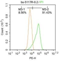
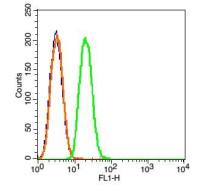
| 产品图片 |  Sample: Kidney (Mouse) Lysate at 40 ug Liver (Mouse) Lysate at 40 ug Primary: Anti-SYVN1 (bs-0679R) at 1/1000 dilution Secondary: IRDye800CW Goat Anti-Rabbit IgG at 1/20000 dilution Predicted band size: 65 kD Observed band size: 65/70 kD  Tissue/cell: mouse kidney tissue; 4% Paraformaldehyde-fixed and paraffin-embedded; Antigen retrieval: citrate buffer ( 0.01M, pH 6.0 ), Boiling bathing for 15min; Block endogenous peroxidase by 3% Hydrogen peroxide for 30min; Blocking buffer (normal goat serum,C-0005) at 37℃ for 20 min; Incubation: Anti-SYVN1/HRD1 Polyclonal Antibody, Unconjugated(bs-0679R) 1:200, overnight at 4°C, followed by conjugation to the secondary antibody(SP-0023) and DAB(C-0010) staining  Tissue/cell: mouse liver tissue; 4% Paraformaldehyde-fixed and paraffin-embedded; Antigen retrieval: citrate buffer ( 0.01M, pH 6.0 ), Boiling bathing for 15min; Block endogenous peroxidase by 3% Hydrogen peroxide for 30min; Blocking buffer (normal goat serum,C-0005) at 37℃ for 20 min; Incubation: Anti-SYVN1/HRD1 Polyclonal Antibody, Unconjugated(bs-0679R) 1:200, overnight at 4°C, followed by conjugation to the secondary antibody(SP-0023) and DAB(C-0010) staining  The figure annotation: The blue histogram is unstained cells. The Wathet Blue histogram is cells stained with secondary antibody (bs-0295G-FITC) alone. The Orange histogram is cells stained with rabbit IgG isotype control antibody(bs-0295P)plus secondary antibody. The green histogram is cells stained with Rabbit Anti-SYVN1 antibody (bs-0679R) plus secondary antibody. Positive control: Hepg2 cells Concentration: 1:50 |


上海联迈生物工程有限公司
品牌商实名认证
钻石会员
入驻年限:8年
