
产品详情
文献和实验
相关推荐
免疫原 :KLH conjugated
亚型 :IgG
形态 :冻干粉
保存条件 :负20°保存
克隆性 :多克隆
标记物 :详情请来电索取说明书
适应物种 :详情请来电索取说明书
宿主 :Rabbit
应用范围 :WB=1:500-2000 ELISA=1:500-1000 IHC-P=1:400-800 IHC-F=1:400-800 IF=1:100-500
浓度 :1mg/ml
靶点 :详情请来电索取说明书
抗体英文名 :CD4
抗体名 :CD4抗体
规格 :100ul
英文名称CD4中文名称CD4抗体
别 名CD4 (L3T4); CD4 antigen (p55); CD4 Antigen ; CD4 molecule; CD4 Receptor; CD4+ Lymphocyte deficiency, included; CD4mut; L3T4; Leu3; Ly-4; Lymphocyte antigen CD4; MGC165891; p55; T Cell Antigen T4 ; T cell antigen T4/LEU3; T cell differentiation antigen L3T4; T cell OKT4 deficiency, included; T cell surface antigen T4/Leu 3 ; T cell surface antigen T4/Leu3; T Cell Surface Glycoprotein CD4; W3/25; W3/25 antigen; T-cell surface glycoprotein CD4 isoform 1 precursor; CD4_HUMAN.
文献引用

规格价格50ul/780元 购买 100ul/1380元 购买 200ul/2200元 购买 大包装/询价
说 明 书50ul 100ul 200ul
研究领域肿瘤 细胞生物 免疫学 干细胞 转录调节因子 细胞表面分子 t-淋巴细胞
抗体来源Rabbit
克隆类型Polyclonal
交叉反应 Human, Mouse, Rat, Dog, Pig, Cow, Sheep, Guinea Pig, MACFA
产品应用WB=1:500-2000 ELISA=1:500-1000 IHC-P=1:400-800 IHC-F=1:400-800 Flow-Cyt=1μg/Test IF=1:100-500 (石蜡切片需做抗原修复)
not yet tested in other applications.
optimal dilutions/concentrations should be determined by the end user.
分 子 量48kDa
细胞定位细胞膜
性 状Lyophilized or Liquid
浓 度1mg/ml
免 疫 原KLH conjugated synthetic peptide derived from human CD4:385-457/457 <Cytoplasmic>
亚 型IgG
纯化方法affinity purified by Protein A
储 存 液0.01M TBS(pH7.4) with 1% BSA, 0.03% Proclin300 and 50% Glycerol.
保存条件Store at -20 °C for one year. Avoid repeated freeze/thaw cycles. The lyophilized antibody is stable at room temperature for at least one month and for greater than a year when kept at -20°C. When reconstituted in sterile pH 7.4 0.01M PBS or diluent of antibody the antibody is stable for at least two weeks at 2-4 °C.
PubMedPubMed
产品介绍background:
This gene encodes a membrane glycoprotein of T lymphocytes that interacts with major histocompatibility complex class II antigenes and is also a receptor for the human immunodeficiency virus. This gene is expressed not only in T lymphocytes, but also in B cells, macrophages, and granulocytes. It is also expressed in specific regions of the brain. The protein functions to initiate or augment the early phase of T-cell activation, and may function as an important mediator of indirect neuronal damage in infectious and immune-mediated diseases of the central nervous system. Multiple alternatively spliced transcript variants encoding different isoforms have been identified in this gene. [provided by RefSeq, Aug 2010].
Function:
Accessory protein for MHC class-II antigen/T-cell receptor interaction. May regulate T-cell activation. Induces the aggregation of lipid rafts.
Subunit:
Associates with LCK. Binds to HIV-1 gp120 and to P4HB/PDI and upon HIV-1 binding to the cell membrane, is part of P4HB/PDI-CD4-CXCR4-gp120 complex. Interacts with HIV-1 Envelope polyprotein gp160 and protein Vpu. Interacts with Human Herpes virus 7 capsid proteins. Interacts with PTK2/FAK1; this interaction requires the presence of HIV-1 gp120.
Subcellular Location:
Cell membrane; Single-pass type I membrane protein. Note=Localizes to lipid rafts. Removed from plasma membrane by HIV-1 Nef protein that increases clathrin-dependent endocytosis of this antigen to target it to lysosomal degradation. Cell surface expression is also down-modulated by HIV-1 Envelope polyprotein gp160 that interacts with, and sequesters CD4 in the endoplasmic reticulum.
Post-translational modifications:
Palmitoylation and association with LCK contribute to the enrichment of CD4 in lipid rafts.
Similarity:
Contains 3 Ig-like C2-type (immunoglobulin-like) domains.
Contains 1 Ig-like V-type (immunoglobulin-like) domain.
SWISS:
P05540
Gene ID:
920
Database links:
Entrez Gene: 920 Human
Entrez Gene: 12504 Mouse
Omim: 186940 Human
SwissProt: P06332 Mouse
SwissProt: P01730 Human
Unigene: 631659 Human
Unigene: 2209 Mouse
Important Note:
This product as supplied is intended for research use only, not for use in human, therapeutic or diagnostic applications.
此抗体可识别55KDⅠ型单链穿膜糖蛋白。 CD4分子是存在于大多数辅助/诱导T细胞表面的59kDa的糖蛋白。正常淋巴组织中CD4的表达数量多于CD8,此抗体主要用于标记辅助/诱导T细胞,与CD8单抗联合使用对外周血淋巴细胞分型。 CD4抗原是HLA-II类分子和人类免疫缺陷病毒(HIV)-爱滋病的受体,在35-50%外周血淋巴细胞-辅助和诱导T细胞(Th/Ti)和70-80%人胸腺细胞上表达,在人的单核细胞表面也有低密度的表达。 CD4抗原有膜结合型和可溶性两种形式。Th/Ti可辅助Ig产生和T细胞毒T细胞的作用。

Sample:
HL60 Cell Lysate at 40 ug
Primary: Anti- CD4 (bs-0647R) at 1/300 dilution
Secondary: IRDye800CW Goat Anti-Rabbit IgG at 1/20000 dilution
Predicted band size: 48 kD
Observed band size: 55kD

U937 Cell Lysate at 40 ug
Primary: Anti- CD4 (bs-0647R) at 1/300 dilution
Secondary: IRDye800CW Goat Anti-Rabbit IgG at 1/20000 dilution
Predicted band size: 48 kD
Observed band size: 55kD

Sample:
Spleen (Mouse) Lysate at 40 ug
Primary: Anti- CD4 (bs-0647R) at 1/300 dilution
Secondary: IRDye800CW Goat Anti-Rabbit IgG at 1/20000 dilution
Predicted band size: 48 kD
Observed band size: 55kD

Sample:
Lymph node(Mouse) Lysate at 40 ug
Primary: Anti- CD4 (bs-0647R) at 1/300 dilution
Secondary: IRDye800CW Goat Anti-Rabbit IgG at 1/20000 dilution
Predicted band size: 48 kD
Observed band size: 55kD

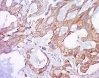
Independently Validated Antibody, image provided by Science Direct, badge number 029634:Formalin-fixed and paraffin embedded human kidney labeled with Anti-CD4 Polyclonal Antibody, Unconjugated (bs-0647R) at 1:400 followed by conjugation to the secondary antibody and DAB staining

Image was kindly submitted by Dr.David M Burmeister from US Army Institute of Surgical Research. Pig lymph nodes stained with Rabbit Anti-CD4 Polyclonal Antibody(bs-bs-0647R)at 1:300 for one hour at room temperature.

Blank control: Mouse splen.
Primary Antibody (green line): Rabbit Anti-CD4 antibody (bs-0647R-FITC)
Dilution: 3μg /10^6 cells;
Isotype Control Antibody (orange line): Rabbit IgG .
Dilution: 3μg /test.
Protocol
The cells incubated in 5%BSA to block non-specific protein-protein interactions for 30 min at room temperature .Cells stained with Primary Antibody for 30 min at room temperature. Acquisition of 20,000 events was performed.

Blank control (Black line): Molt4 (Black). Primary Antibody (green line): Rabbit Anti-CD4 antibody (bs-0647R-PE) Dilution: 3μg /10^6 cells; Isotype Control Antibody (orange line): Rabbit IgG-PE. Protocol The cells were fixed with 4% PFA (10min at room temperature)and then were incubated in 5%BSA to block non-specific protein-protein interactions for 30 min at room temperature .Cells stained with Primary Antibody for 30 min at room temperature. Acquisition of 20,000 events was performed.

Blank control (blue line): HL60 (fixed with 2% paraformaldehyde (10 min) and then permeabilized with 0.1% PBS-Tween for 20 min at room temperature).
Primary Antibody (green line): Rabbit Anti-CD4 antibody (bs-0647R),Dilution: 1μg /10^6 cells;
Isotype Control Antibody (orange line): Rabbit IgG .
Secondary Antibody (white blue line): Goat anti-rabbit IgG-FITC,dilution: 1μg /test.


上海雅吉生物科技有限公司
品牌商实名认证
皇冠会员
入驻年限:8年
