
产品详情
文献和实验
相关推荐
免疫原 :KLH conjugated synthetic(具体详情咨询我们)
亚型 :IgG
形态 :Lyophilized or Liquid
保存条件 :Store at -20 °C
克隆性 :多克隆
标记物 :有各种标记抗体需要可以联系我公司
适应物种 :Hu Mo Pig Cow Hor Sheep等
宿主 :Rabbit
应用范围 :WB=1:100-500,Elisa=1:500-1000,IHC-P=1:100-500,IHC-F=1:100-500,Flow Cyt=1μg |test,IF=1:100-500,ICC=1:100-2409,
浓度 :1mg/ml
靶点 :详询
抗体英文名 :Vimentin
抗体名 :波形蛋白抗体
规格 :100ul
英文名称Vimentin中文名称波形蛋白抗体
别 名VIM; FLJ36605; OTTHUMP00000019224; VIM; VIME_HUMAN; Vimentin.
Specific References (4) | bs-8533R has been referenced in 4 publications.[IF=2.52] Yang, Ning, et al. "Overexpression of SOX2 promotes migration, invasion, and epithelial-mesenchymal transition through the Wnt/β-catenin pathway in laryngeal cancer Hep-2 cells." Tumor Biology (2014): 1-9. WB ; Human. PubMed:24833089 [IF=4.14] He, D., et al. "Enhanced M1/M2 Macrophage Ratio Promotes Orthodontic Root Resorption." Journal of Dental Research (2014): 0022034514553817. Rat. PubMed:25344334 [IF=4.42] Gao, Lili, et al. "Glycyrrhizic acid alleviates bleomycin-induced pulmonary fibrosis in rats." Frontiers in pharmacology 6 (2015). WB ; Rat. PubMed:26483688 [IF=1.56] Huang, Chao, et al. "Analysis of different components in the peritumoral tissue microenvironment of colorectal cancer: A potential prospect in tumorigenesis. Corrigendum in/10.3892/mmr. 2016.5882." Molecular Medicine Reports 14.3 (2016): 2555-2565. IHC-P ; Human. PubMed:27484148
规格100ul 200ul
研究领域肿瘤 细胞生物 免疫学 信号转导 干细胞 细胞骨架 肿瘤细胞生物标志物
抗体来源Rabbit
克隆类型Polyclonal
交叉反应 Human, Mouse, Rat, Dog, Pig, Cow, Horse, Rabbit,
产品应用WB=1:500-2000 ELISA=1:500-1000 IHC-P=1:400-800 IHC-F=1:400-800 Flow-Cyt=1μg/Test ICC=1:100-500 IF=1:100-500 (石蜡切片需做抗原修复)
not yet tested in other applications.
optimal dilutions/concentrations should be determined by the end user.
分 子 量51kDa
细胞定位细胞浆
性 状Lyophilized or Liquid
浓 度1mg/ml
免 疫 原KLH conjugated synthetic peptide derived from human Vimentin:371-466/466
亚 型IgG
纯化方法affinity purified by Protein A
储 存 液0.01M TBS(pH7.4) with 1% BSA, 0.03% Proclin300 and 50% Glycerol.
保存条件Store at -20 °C for one year. Avoid repeated freeze/thaw cycles. The lyophilized antibody is stable at room temperature for at least one month and for greater than a year when kept at -20°C. When reconstituted in sterile pH 7.4 0.01M PBS or diluent of antibody the antibody is stable for at least two weeks at 2-4 °C.
PubMedPubMed
产品介绍background:
This gene encodes a member of the intermediate filament family. Intermediate filamentents, along with microtubules and actin microfilaments, make up the cytoskeleton. The protein encoded by this gene is responsible for maintaining cell shape, integrity of the cytoplasm, and stabilizing cytoskeletal interactions. It is also involved in the immune response, and controls the transport of low-density lipoprotein (LDL)-derived cholesterol from a lysosome to the site of esterification. It functions as an organizer of a number of critical proteins involved in attachment, migration, and cell signaling. Mutations in this gene causes a dominant, pulverulent cataract.[provided by RefSeq, Jun 2009]
Function:
Vimentins are class-III intermediate filaments found in various non-epithelial cells, especially mesenchymal cells. Vimentin is attached to the nucleus, endoplasmic reticulum, and mitochondria, either laterally or terminally. Involved with LARP6 in the stabilization of type I collagen mRNAs for CO1A1 and CO1A2. Subunit : Homopolymer assembled from elementary dimers. Interacts with HCV core protein. Interacts with LGSN and SYNM. Interacts (via rod region) with PLEC (via CH 1 domain) (By similarity). Interacts with SLC6A4. Interacts with STK33. Interacts with LARP6. Interacts with RAB8B (By similarity).
Subcellular Location:
Cytoplasm.
Tissue Specificity:
Highly expressed in fibroblasts, some expression in T- and B-lymphocytes, and little or no expression in Burkitt's lymphoma cell lines. Expressed in many hormone-independent mammary carcinoma cell lines.
Post-translational modifications:
Filament disassembly during mitosis is promoted by phosphorylation at Ser-55 as well as by nestin. One of the most prominent phosphoproteins in various cells of mesenchymal origin. Phosphorylation is enhanced during cell division, at which time vimentin filaments are significantly reorganized. Phosphorylation by PKN1 inhibits the formation of filaments. Phosphorylated at Ser-56 by CDK5 during neutrophil secretion in the cytoplasm. Phosphorylated by STK33.
Similarity:
Belongs to the intermediate filament family.
SWISS:
P08670
Gene ID:
7431
Database links:
Entrez Gene: 7431 Human
Entrez Gene: 22352 Mouse
Entrez Gene: 81818 Rat
Omim: 193060 Human
SwissProt: P08670 Human
SwissProt: P20152 Mouse
SwissProt: P31000 Rat
Unigene: 455493 Human
Important Note:
This product as supplied is intended for research use only, not for use in human, therapeutic or diagnostic applications.
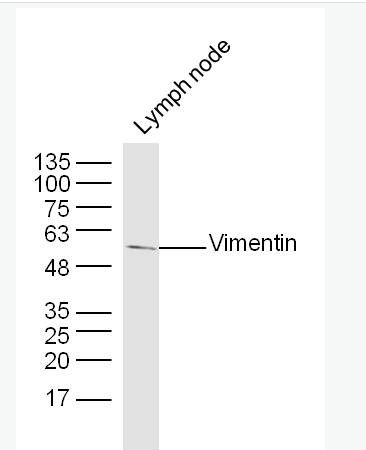
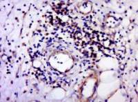
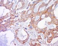
| 产品图片 | 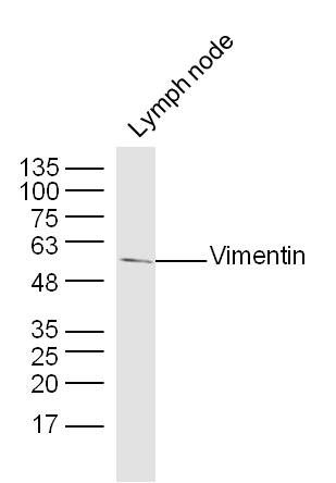 Sample: lymph node (Mouse) Lysate at 40 ug Primary: Anti-Vimentin(bs-8533R) at 1/300 dilution Secondary: IRDye800CW Goat Anti-Rabbit IgG at 1/20000 dilution Predicted band size: 51 kD Observed band size: 51 kD 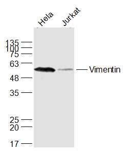 Sample: A549(Human) Cell Lysate at 30 ug Jurkat(Human) Cell Lysate at 30 ug Primary: Anti-Vimentin (bs-8533R) at 1/1000 dilution Secondary: IRDye800CW Goat Anti-Rabbit IgG at 1/20000 dilution Predicted band size: 51 kD Observed band size: 53 kD 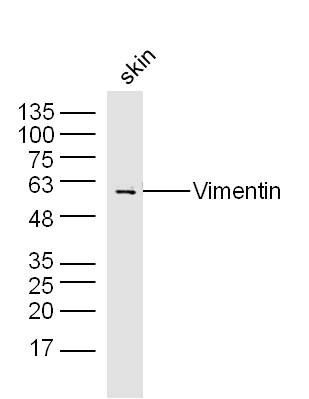 Sample: skin(Mouse) Lysate at 40 ug Primary: Anti-Vimentin(bs-8533R) at 1/300 dilution Secondary: IRDye800CW Goat Anti-Rabbit IgG at 1/20000 dilution Predicted band size: 51 kD Observed band size: 51 kD 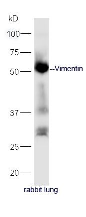 Protein: lung(rabbit) lysate at 40ug; Primary: rabbit Anti-Vimentin (bs-8533R) at 1:300; Secondary: HRP conjugated Goat-Anti-rabbit IgG(bs-0295G-HRP) at 1: 5000; Predicted band size: 51 kD Observed band size: 51 kD 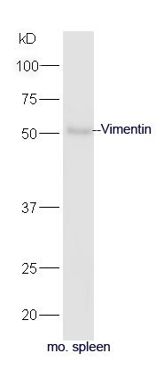 Protein: spleen(mouse) lysate at 40ug; Primary: rabbit Anti-Vimentin (bs-8533R) at 1:300; Secondary: HRP conjugated Goat-Anti-rabbit IgG(bs-0295G-HRP) at 1: 5000; Predicted band size: 51 kD Observed band size: 51 kD 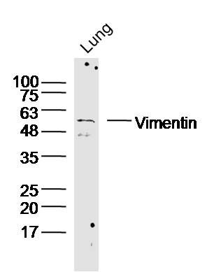 Sample: Lung (Mouse) Lysate at 40 ug Primary: Anti-Vimentin (bs-8533R) at 1/300 dilution Secondary: IRDye800CW Goat Anti-Rabbit IgG at 1/20000 dilution Predicted band size: 51kD Observed band size: 51kD 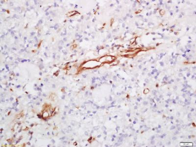 Tissue/cell: human lung carcinoma; 4% Paraformaldehyde-fixed and paraffin-embedded; Antigen retrieval: citrate buffer ( 0.01M, pH 6.0 ), Boiling bathing for 15min; Block endogenous peroxidase by 3% Hydrogen peroxide for 30min; Blocking buffer (normal goat serum,C-0005) at 37∩ for 20 min; Incubation: Anti-Vimentin Polyclonal Antibody, Unconjugated(bs-8533R) 1:200, overnight at 4∑C, followed by conjugation to the secondary antibody(SP-0023) and DAB(C-0010) staining 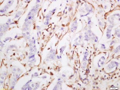 Tissue/cell: human breast carcinoma; 4% Paraformaldehyde-fixed and paraffin-embedded; Antigen retrieval: citrate buffer ( 0.01M, pH 6.0 ), Boiling bathing for 15min; Block endogenous peroxidase by 3% Hydrogen peroxide for 30min; Blocking buffer (normal goat serum,C-0005) at 37∩ for 20 min; Incubation: Anti-Vimentin Polyclonal Antibody, Unconjugated(bs-8533R) 1:200, overnight at 4∑C, followed by conjugation to the secondary antibody(SP-0023) and DAB(C-0010) staining 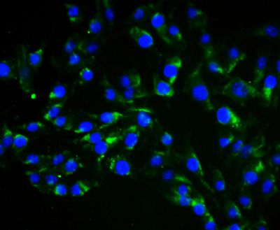 Tissue/cell: endothelial cells of umbilical artery;4% Paraformaldehyde-fixed; Blocking buffer (normal goat serum,C-0005) at 37℃ for 20 min; Incubation: Anti-Vimentin Polyclonal Antibody, Alexa Fluor 488 conjugated(bs-8533R-A488) 1:100, 60 minutes at 37℃. DAPI(5ug/ml,blue,C-0033) was used to stain the cell nuclei 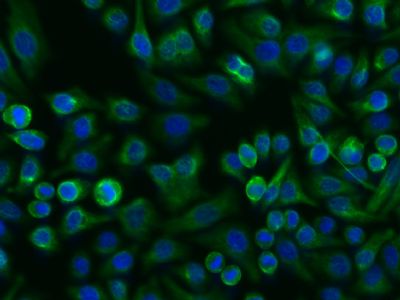 Tissue/cell: FHC cell; 4% Paraformaldehyde-fixed; Triton X-100 at room temperature for 20 min; Blocking buffer (normal goat serum, C-0005) at 37°C for 20 min; Antibody incubation with (Vimentin) Polyclonal Antibody, Unconjugated (bs-8533R) 1:200, 2 hours at 37°C; followed by a conjugated Goat Anti-Rabbit IgG antibody (bs-0295G-FITC) at 37°C for 90 minutes, DAPI (5ug/ml, blue, C-0033) was used to stain the cell nuclei. 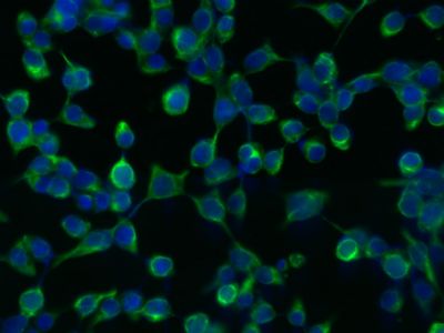 Tissue/cell: 293T cell; 4% Paraformaldehyde-fixed; Triton X-100 at room temperature for 20 min; Blocking buffer (normal goat serum, C-0005) at 37°C for 20 min; Antibody incubation with (Vimentin) Polyclonal Antibody, Unconjugated (bs-8533R) 1:200, 2 hours at 37°C; followed by a conjugated Goat Anti-Rabbit IgG antibody (bs-0295G-FITC) at 37°C for 90 minutes, DAPI (5ug/ml, blue, C-0033) was used to stain the cell nuclei. 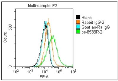 Blank control (blue line): U251(blue). Primary Antibody (green line): Rabbit Anti-Vimentin antibody (bs-8533R) Dilution: 2μg /10^6 cells; Isotype Control Antibody (orange line): Rabbit IgG . Secondary Antibody (white blue line): Goat anti-rabbit IgG-PE Dilution: 1μg /test. Protocol The cells were fixed with 4% paraformaldehyde (10 min) and then permeabilized with 0.1% PBS-Tween for 20 min at room temperature Cells stained with Primary Antibody for 30 min at room temperature. The cells were then incubated in 1 X PBS/2%BSA/10% goat serum to block non-specific protein-protein interactions followed by the antibody for 15 min at room temperature. The secondary antibody used for 40 min at room temperature. Acquisition of 20,000 events was performed. |


上海雅吉生物科技有限公司
品牌商实名认证
皇冠会员
入驻年限:8年
