
产品详情
文献和实验
相关推荐
免疫原 :Carrier-protein conjugated synthetic peptide encompassing a sequence within the C-terminus region of human Iba1. The exact sequence is proprietary.
亚型 :IgG
形态 :Liquid
保存条件 :Store as concentrated solution. Centrifuge briefly prior to opening vial. For short-term storage (1-2 weeks), store at 4ºC. For long-term storage, aliquot and store at -20ºC or below. Avoid multiple freeze-thaw cycles.
克隆性 :Polyclonal
标记物 :Unconjugated
适应物种 :Human, Mouse, Rat
保质期 :12 months from the shipping date of the product.
抗原来源 :Human
目录编号 :GTX100042
级别 :Primary Antibodies
库存 :Available
供应商 :GeneTex
宿主 :Rabbit
应用范围 :WB, ICC/IF, IHC-P, IHC-Fr, FACS, IHC, IHC (Free Floating)
浓度 :0.06 mg/ml (Please refer to the vial label for the specific concentration.)
靶点 :Iba1
抗体英文名 :Iba1 antibody
抗体名 :Iba1 抗体
规格 :100 μl/25 μl
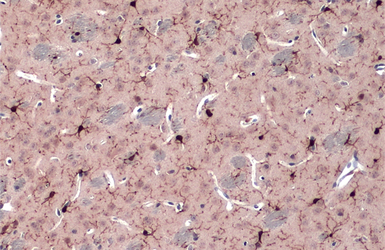
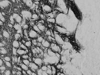
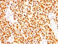
Iba1 antibody detects Iba1 protein by immunohistochemical analysis.Sample: Paraffin-embedded rat tissues.Iba1 stained by Iba1 antibody (GTX100042) diluted at 1:100.Antigen Retrieval: Citrate buffer, pH 6.0, 15 min
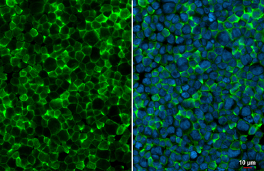
Iba1 antibody detects Iba1 protein at cell membrane by immunofluorescent analysis.Sample: THP-1 cells were fixed in 4% PFA at RT for 15 min.Green: Iba1 stained by Iba1 antibody (GTX100042) diluted at 1:500.Blue: Fluoroshield with DAPI (GTX30920).
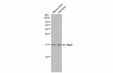
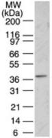
Various tissue extracts (50 μg) were separated by 15% SDS-PAGE, and the membrane was blotted with Iba1 antibody (GTX100042) diluted at 1:1000. The HRP-conjugated anti-rabbit IgG antibody (GTX213110-01) was used to detect the primary antibody.
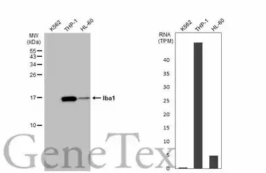
Various whole cell extracts (30 μg) were separated by 15% SDS-PAGE, and the membrane was blotted with Iba1 antibody (GTX100042) diluted at 1:5000. The HRP-conjugated anti-rabbit IgG antibody (GTX213110-01) was used to detect the primary antibody. Correspo
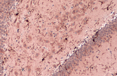
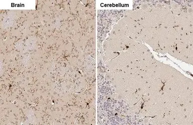
Iba1 antibody detects Iba1 protein at cell membrane and cytoplasm by immunohistochemical analysis.Sample: Paraffin-embedded rat brain.Iba1 stained by Iba1 antibody (GTX100042) diluted at 1:500.Antigen Retrieval: Citrate buffer, pH 6.0, 15 min
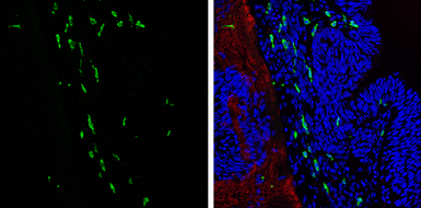
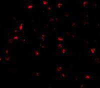
Iba1 antibody detects Iba1 protein expression at microglias by immunohistochemical analysis.
Sample: Frozen sectioned E13.5 Rat brain.
Green: Iba1 protein stained by Iba1 antibody (GTX100042) diluted at 1:250.
Red: beta Tubulin 3/ TUJ1, a mature neuron marker, stained by beta Tubulin 3/ TUJ1 antibody [GT11710] (GTX631836) diluted at 1:500.
Blue: Fluoroshield with DAPI (GTX30920).

Iba1 antibody detects Iba1 protein at cell membrane and cytoplasm by immunohistochemical analysis.Sample: Frozen-sectioned mouse brain.Green: Iba1 stained by Iba1 antibody (GTX100042) diluted at 1:500.Blue: Fluoroshield with DAPI (GTX30920).Antigen Retrieval: ice-cold MeOH for 5 min
Iba1 antibody detects Iba1 protein at cell membrane and cytoplasm by immunohistochemical analysis.Sample: Paraffin-embedded mouse brain.Iba1 stained by Iba1 antibody (GTX100042) diluted at 1:500.Antigen Retrieval: Citrate buffer, pH 6.0, 15 min

Rat tissue extract (50 μg) was separated by 15% SDS-PAGE, and the membrane was blotted with Iba1 antibody (GTX100042) diluted at 1:1000.

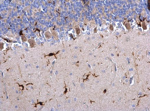
Iba1 antibody detects Iba1 protein on rat hind brain by immunohistochemical analysis.
Sample: Paraffin-embedded rat hind brain.
Iba1 antibody (GTX100042) dilution: 1:500.
Antigen Retrieval: Trilogy™ (EDTA based, pH 8.0) buffer, 15min
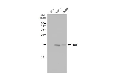
Various whole cell extracts (30 μg) were separated by 15% SDS-PAGE, and the membrane was blotted with Iba1 antibody (GTX100042) diluted at 1:5000. The HRP-conjugated anti-rabbit IgG antibody (GTX213110-01) was used to detect the primary antibody.


GeneTex
品牌商实名认证
钻石会员
入驻年限:12年
