
产品详情
文献和实验
相关推荐
库存 :99
供应商 :biocyto
肿瘤类型 :CN
细胞类型 :Adherent
ATCC Number :NC
品系 :NC
组织来源 :Kindy
相关疾病 :NC
物种来源 :Rat/Mouse/Human
免疫类型 :NC
是否是肿瘤细胞 :否
器官来源 :Kindy
运输方式 :Liquid Nitrogen
年限 :6个月
生长状态 :存活率95%以上
规格 :10^6
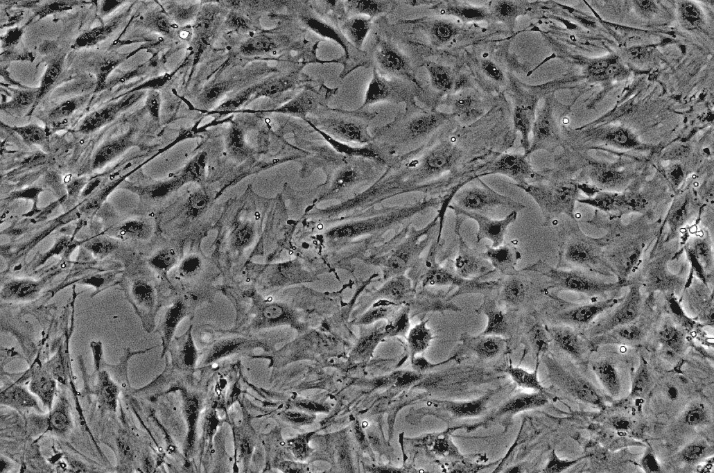
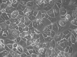
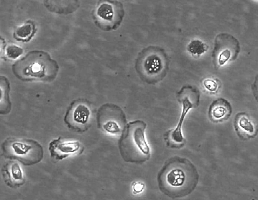
DescriptionRenal mesangial cells (RMC) are perivascular pericytes located within the central portion of the glomerular tuft between capillary loops. Mesangial cells have a variety of functions including synthesis and assembly of the mesangial matrix, endocytosis and processing of plasma macromolecules, and control of glomerular hemodynamics via mesangial cell contraction or release of vasoactive hormones. Mesangial cell proliferation and matrix overproduction are the predominant pathological features of many forms of glomerulonephritis, such as IgA nephropathy, lupus nephropathy, and diabetic nephropathy and frequently precedes the increase of extracellular matrix in the mesangium and the development of glomerulosclerosis . As proliferation and matrix synthesis/degradation in vitro are regulated by cytokines and growth factors, cultured cells are an ideal tool for studying pathophysiological events such as mesangial expansion, scarring, and glomerulosclerosis.
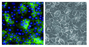
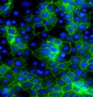
HRMC from ScienCell Research Laboratories are isolated from human renal tissue. HRMC are cryopreserved immediately after purification and delivered frozen. Each vial contains >5 x 10⁵ cells in 1 ml volume. HRMC are characterized by immunofluorescent method with antibodies to fibronectin, Thy-1, and smooth muscle actin. HRMC are negative for HIV-1, HBV, HCV, mycoplasma, bacteria, yeast and fungi. HBMEC are guaranteed to further expand for 15 population doublings in the conditions specified by ScienCell Research Laboratories.
| Product | Catalog no. | Amount | Storage |
| Renal mesangial cells | RMC001 | 1X10^6/vial | in liquid nitrogen |
Product Use
For Research Use Only.Not for use in diagnostic procedures
Culture Conditions
Culture Type:Adherent
Temperature Range:36℃ to 38℃
Incubator Atmosphere:Humidified atmosphere of 5% CO2
Passaging Adherent Cells
All solutions and equipment that come in contact with the cells must be sterile.
Always use proper sterile technique and work in a laminar flow hood.
1. Remove and discard the spent cell culture media from the culture vessel.
2. Wash cells using a balanced salt solution without calcium and magnesium (approximately 2 mL per 10 cm2 culture surface area). Gently add washsolution to the side of the vessel opposite the attached cell layer to avoiddisturbing the cell layer,and rock the vessel back and forth several times.
Note: The wash step removes any traces of serum, calcium, and magnesium that would inhibit the action of the dissociation reagent.
3. Remove and discard the wash solution from the culture vessel
4. Add the pre-warmed dissociation reagent such as trypsin or TrypLE™to the side of the flask; use enough reagent to cover the cell layer (approximately0.5 mL per 10 cm2). Gently rock the container to get complete coverage of the cell layer.
5. Incubate the culture vessel at room temperature for approximately 2minutes.
Note: that the actual incubation time varies with the cell line used.
6. Observe the cells under the microscope for detachment. If cells areless than 90% detached, increase the incubation time a few more minutes, checking for dissociation every 30 seconds. You may also tap the vessel to expedite cell Tetachment.
7. When ≥ 90% of the cells have detached, tilt the vessel for a minimallength of time to allow the cells to drain. Add the equivalent of 2 volumes (twice thevolume used for the dissociation reagent) of pre-warmed complete growth medium.Disperse the medium by pipetting over the cell layer surface several times.
8. Transfer the cells to a 15-mL conical tube and centrifuge then at200 × g for 5 to 10 minutes. Note that the centrifuge speed and time vary based on the cell type.
9. Resuspend the cell pellet in a minimal volume of pre-warmed complete growth medium and remove a sample for counting.
10. Determine the total number of cells and percent viability using a hemacytometer, cell counter and Trypan Blue exclusion, or the Countess® Automated CellCounter. If necessary, add growth media to the cells to achieve the desired cellconcentration and recount the cells.


广州柏赛柯生物技术有限公司
代理商实名认证
钻石会员
入驻年限:8年
