
产品详情
文献和实验
相关推荐
ATCC Number :CRL-2142
库存 :现货
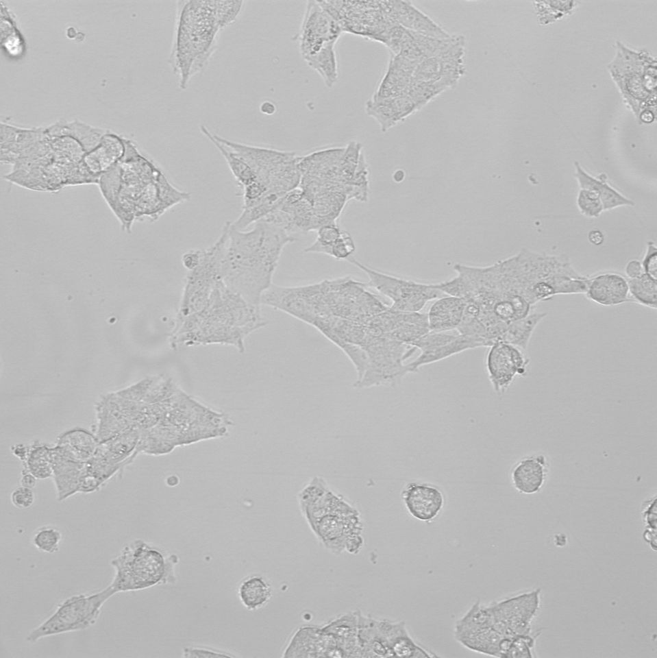
生长状态 :贴壁生长
运输方式 :常温/干冰
器官来源 :脑
是否是肿瘤细胞 :是
细胞形态 :上皮细胞样
物种来源 :人
规格 :1×10^6 cells
SK-N-FI
SK-N-FI are neuroblasts that were isolated from the brain of an 11-year-old, White, male patient with neuroblastoma. It has applications in immunology and neuroscience.
Product category |
Human cells |
Synonyms |
SK_N_FI; SKNFI; SK-N-F1; SKNF1 |
Organism |
Homo sapiens, human |
Cell type |
neuroblast |
Morphology |
epithelial |
Tissue |
Brain |
Disease |
Neuroblastoma |
Applications |
3D cell culture Immunology Neuroscience |
Characteristics
Growth properties |
Adherent |
Derivation |
SK-N-FI is a neuroblastoma cell line derived in 1979 from a bone marrow metastasis from a 11 year old Caucasian male with poorly differentiated embryonal neuroblastoma. |
Population doubling time |
- |
Age |
11 years |
Ethnicity |
White |
Gender |
Male |
Tumorigenic |
Yes; Yes, forms tumors in nude mice with a latent period of 4 to 6 weeks |
Metastatic |
Bone marrow |
Genes expressed |
tumor necrosis factor (TNF) |
Expression markers |
Tumor necrosis factor (TNF) type B, expressed; tumor necrosis factor (TNF) type A, expressed |
Comments |
The cells exhibit high MDR1 expression. Recombinant tumor necrosis factor (TNF) stimulates the proliferation of SK-N-FI cells in both serum free medium and fetal bovine serum supplemented medium, but has no effect in medium without insulin. |
Handling information
Unpacking and storage instructions |
1. Check all containers for leakage or breakage. 2. Remove the frozen cells from the dry ice packaging and immediately place the cells at a temperature below -130°C, preferably in liquid nitrogen vapor, until ready for use. |
Complete medium |
The base medium for this cell line is ATCC-formulated Dulbecco's Modified Eagle's Medium, Catalog No. 30-2002. To make the complete growth medium, add the following components to the base medium: l 0.1 mM Non-Essential Amino Acids (NEAA) l fetal bovine serum to a final concentration of 10% |
Temperature |
37°C |
Atmosphere |
95% Air, 5% CO2 |
Handling procedure |
To insure the highest level of viability, thaw the vial and initiate the culture as soon as possible upon receipt. If upon arrival, continued storage of the frozen culture is necessary, it should be stored in liquid nitrogen vapor phase and not at -70°C. Storage at -70°C will result in loss of viability. 1. Thaw the vial by gentle agitation in a 37°C water bath. To reduce the possibility of contamination, keep the O-ring and cap out of the water. Thawing should be rapid (approximately 2 minutes). 2. Remove the vial from the water bath as soon as the contents are thawed, and decontaminate by dipping in or spraying with 70% ethanol. All of the operations from this point on should be carried out under strict aseptic conditions. 3. Transfer the vial contents to a 75 cm2 tissue culture flask and dilute with the recommended complete culture medium (see the specific batch information for the recommended dilution ratio). It is important to avoid excessive alkalinity of the medium during recovery of the cells. It is suggested that, prior to the addition of the vial contents, the culture vessel containing the complete growth medium be placed into the incubator for at least 15 minutes to allow the medium to reach its normal pH (7.0 to 7.6). 4. Incubate the culture at 37°C in a suitable incubator. A 5% CO2 in air atmosphere is recommended if using the medium described on this product sheet. Note: It is not necessary to remove the cryoprotective agent. If it is desired that the cryoprotective agent be removed immediately, or that a more concentrated cell suspension be obtained, centrifuge the cell suspension at approximately 125 x g for 5 to 10 minutes. Discard the supernatant and resuspend the cells with fresh growth medium at the dilution ratio recommended in the specific batch information. |
Subculturing procedure |
Volumes used in this protocol are for 75 cm2 flask; proportionally reduce or increase amount of dissociation medium for culture vessels of other sizes. 1. Remove and discard culture medium. 2. Briefly rinse the cell layer with 0.25% (w/v) Trypsin-0.53 mM EDTA solution to remove all traces of serum that contains trypsin inhibitor. 3. Add 2.0 to 3.0 mL of Trypsin-EDTA solution to flask and ob-serve cells under an inverted microscope until cell layer is dispersed (usually within 5 to 15 minutes). Note: To avoid clumping do not agitate the cells by hitting or shaking the flask while waiting for the cells to detach. Cells that are difficult to detach may be placed at 37°C to facilitate dispersal. 4. Add 6.0 to 8.0 mL of complete growth medium and aspirate cells by gently pipetting. 5. Add appropriate aliquots of the cell suspension to new culture vessels. 6. Incubate cultures at 37°C. Subculture Ratio: 1:4 Medium Renewal: 2 to 3 times a week. |
Reagents for cryopreservation |
Complete growth medium supplemented with 5% (v/v) DMSO. |
Quality control specifications
Mycoplasma contamination |
Not detected |
STR profiling |
Amelogenin: X,Y CSF1PO: 10,12 D13S317: 11,14 D16S539: 11,12 D5S818: 12,13 D7S820: 8,9 TH01: 6,9.3 TPOX: 8,9 vWA: 16,17 D3S1358: 16,18 D21S11: 28,30 D18S51: 12,14 Penta_E: 11,20 Penta_D: 2.2,13 D8S1179: 11,12 FGA: 22 D19S433: 12 D2S1338: 17,18 |


上海赛澜斯生物技术有限责任公司
实名认证
金牌会员
入驻年限:1年
